Download
1 / 1
10 likes | 74 Views
3D ultrasound and 4D ultrasound are performed in the same way as a 2D doctor's ultrasound: the mother's belly is coated with gel, and then a probe is passed over. The ultrasound is the same as in a 2D ultrasound, only the scanning method changes, allowing to see a baby in three dimensions. <br>

E N D
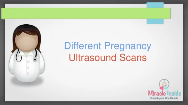
Different types of Ultrasound Test Prevalent these Days 3D ultrasound and 4D ultrasound are performed in the same way as a 2D doctor's ultrasound: the mother's belly is coated with gel, and then a probe is passed over. The ultrasound is the same as in a 2D ultrasound, only the scanning method changes, allowing to see a baby in three dimensions. Thanks to a 3D probe coupled to an image reconstruction system, it is the external appearance of Baby that you will be able to visualize. In complicated pregnancies, some women may have ultrasounds every week at their doctor's office without any problem, either for the mother or the child. Before starting the 3D ultrasound, you will have all the necessary explanations on the images that will be shown to you to take full advantage of this magnificent moment. When you come to ultrasound clinic in noida, after your 22nd week of pregnancy, you have already had your morphological ultrasound, so there is no risk of discovering a malformation on the child. Ultrasound is a diagnostic method in which ultrasound is used to draw the inside of the body. During pregnancy is the method used because not emitting radiation is safe for the baby. Today, three ultrasounds are considered essential throughout pregnancy, but four would be advisable. First Ultrasound- It is performed between week 4/12, and the intravaginal is recommended because it is safer and allows a more accurate visualization. It helps determine the date of delivery and control the true evolution of pregnancy. In this ultrasound, cardiac communication, ossification of the nasal cartilage, nuchal fold in case of malformations and other parameters are observed. Second Ultrasound- Performed between week 12/20. In this ultrasound, it is observed if there are malformations and if the development of the fetus follows its normal course. It is recommended to observe the uterine arteries around week 12 to prevent the risk of preeclampsia (hypertension). Third Ultrasound- Performed between the week 20/30. Ratifies that the development is normal. This ultrasound shows the fetal position and the maturation of the placenta. Fourth Ultrasound.- Performed between the 30/40 week. In this ultrasound, the fetal position is checked for delivery, and weight and height are analyzed. 3D ultrasounds- These ultrasounds have the advantage of allowing to see the appearance of the face and the cutaneous surface of the fetus. It is becoming a widespread technique, and Social Security offers it whenever you have the necessary ultrasound. This type of ultrasound helps to detect the cleft lip that is very difficult to observe in normal ultrasound scans. On the other hand, it serves to know the baby because you can see how he laughs, opens his eyes. Finally, we can offer the 4D ultrasound that, unlike the 3D, allows us to have these volumetric images in real time and in movement, with which we can make videos. 4d ultrasound test price in Noidais higher than other previous versions. Both in the 3D and 4D ultrasounds, the soft parts of the baby's body are observed or highlighted, thus obtaining a more transparent image of the baby, which even allows seeing features and characteristics of their facial expressions.