Download
1 / 45
480 likes | 852 Views
Ch. 6. Antigen-antibody interactions: Principles and applications Strength of Ag-Ab interactions - Affinity is a quantitative measure of binding strength - Avidity incorporates affinity of multiple binding sites . Assays detect presence of antigen or antibody diagnosis

E N D
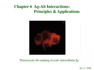
Ch. 6. Antigen-antibody interactions: Principles and applications Strength of Ag-Ab interactions - Affinity is a quantitative measure of binding strength - Avidity incorporates affinity of multiple binding sites Ch. 6
Assays detect presence of antigen or antibody diagnosis monitoring humoral immune response therapeutics analysis of interesting molecules If looking for Ag, use known Ab If looking for Ab, use known Ag Ch. 6
p. 146 Ch. 6
Affinity- strength of interaction between one Ag-binding site and one epitope Measured by various methods including equilibrium dialysis competition assays microchips Association constants can be calculated Ch. 6
Ab avidity Involves multiple interactions between antibody and antigen Both Ab and Ag must be bivalent or multivalent IgM (with 10 antigen-binding sites per molecule): low affinity but high avidity Avidity may be a more biologically significant measure than affinity Ch. 6
Cross-reactivity p. 149 Ch. 6
Precipitation reactions - classic demonstration of antibody-antigen interaction Antibody and soluble antigen form a lattice that eventually develops into a visible precipitate Antibody must be bivalent (monovalent Fabs won’t work) Antigen must be bivalent or multivalent Ch. 6
p. 142, 5th ed. Ch. 6
Precipitation reactions in fluids (p. 142, 5th ed.) p. 150 for SPR app. Precipitation reaction can be seen Ch. 6
Precipitation can also be seen in gels Antibody and antigen diffuse toward each other and precipitate where there is equivalence Radial immunodiffusion – Ab in gel Double diffusion – Ab and Ag diffuse Immunoelectrophoresis - Electrophoresis first - Then precipitation in gel Ch. 6
Ch. 6 p. 152
p. 153 Ch. 6
p. 152 Ch. 6
Agglutination- interaction between antibody and a particulate antigen resulting in visible clumping Presence of excess antibody can inhibit agglutination (prozone effect) - Each antibody “competes” for epitopes - Some antibodies bind but do not agglutinate - Epitope density or availability Ch. 6
p. 153 Ch. 6
Bacterial agglutination to diagnose infection If a patient has a bacterial infection, the patient will produce specific antibodies to that bacterium Serum can be titered with bacterial agglutination reactions Dilutions of serum are tested (usually twofold) example: 1:256 dilution shows agglutination but 1:512 does not Titer is 256 Ch. 6
Titer can be monitored over time; Convalescent serum will have higher titer than acute one (“Paired serum samples”) Antisera are used to type bacteria, too (against surface antigens, flagellar antigens, etc.) Example: E. coli O157:H7 Ch. 6
Passive agglutination Useful with soluble Ag’s that don’t agglutinate But if you stick them onto something else (like a latex bead or, historically, a RBC) you can obtain an agglutination reaction Using synthetic beads increases sensitivity Ch. 6
Agglutination inhibition assays Can test for the presence of substances in fluids (e.g., HCG or drugs in urine) Rubella test is an agglutination inhibition assay; rubella virus causes hemagglutination Ab in person’s serum inhibits hemagglutination Ch. 6
Agglutination inhibition (or, Hapten inhibition) p. 146, 5th ed. Current tests are ELISAs Ch. 6
Principle of radioimmunoassay (RIA): Competitive binding of radiolabelled Ag and unlabelled Ag to a high-affinity Ab Highly sensitive Quantitative Radioactive iodine or tritium used Many variations of the assay have been developed Ch. 6
p. 156 Ch. 6
ELISA- enzyme-linked immunosorbent assay Similar in principle to RIA Nearly as sensitive Cheaper and safer Many detection systems have been developed Many variations of the assay have been developed Ch. 6
All ELISAs use an antibody conjugated with an enzyme that turns a colorless substrate into a colored product Many variations: Direct Indirect Sandwich Competitive Qualitative and quantitative to detect presence of Ag or Ab Ch. 6
p. 157 Ch. 6
Chemiluminescence Most sensitive assay available! Luminol, hydrogen peroxide and horseradish peroxidase react to emit light Ab.HRP + Ag Ab.HRP.Ag (luminol, H2O2) light Expose photographic film or luminometer Ch. 6
p. 158 Ch. 6
ELISPOT Ch. 6
Western blotting p. 159 Ch. 6
p. 160 Ch. 6
Immunofluorescence: to visualize Ag on or within cells Antibodies can be labeled with fluorescent dye Can localize binding sites on cells Dyes: Fluorescein, rhodamine, phycoerythrin can be conjugated to Fc region of Ab (so antigen binding is unaffected) Absorb light at one wavelength (UV) and emit light at another wavelength (e.g., red or green) Ch. 6
Immunofluorescence p. 161 Ch. 6
p. 166 Ch. 6
Versatile technique: - Differentiate T cell subsets - Detect Ag-Ab complexes - Localize of target molecules in tissue (variation: immunohistochemical staining) Ch. 6
Flow cytometry is quantitative FACS- fluoresence-activated cell sorter Analyze cell populations Sort cells with different features into different containers (e.g., T and B cells; cells that are producing a cell-surface marker vs. those that are not) Ch. 6
p. 162 Ch. 6
p. 163 Ch. 6
Uses for flow cytometry: Percentage of a total population of cells Measuring antigen density within a population of cells Multiple antibodies can be used to assess several cell surface antigens simultaneously Clinical analysis (leukemia typing) Ch. 6
Immunoelectron microscopy p. 164 Ch. 6
Summary The structure of antibodies enables them to recognize and bind antigen and to perform appropriate effector functions The exquisite specificity and effector activity of antibodies makes them very useful in research and diagnostics Ch. 6
The organization of immunoglobulin genes allows for the formation of over 10 billion antigen specificities Various in vivo and in vitro experimental systems have provided significant insights into the immune response and its regulation Ch. 6