Download
1 / 39
580 likes | 2.31k Views
Trauma during birth. Prepared by : Ayda khader. Feb.2018. Outline. Objective Introduction Trauma to skin and superficial tissue Muscle trauma Nerve trauma Fracture Haemorrhage due to trauma Haemorrhage due to disruption in blood flow Haemorrhage related to coagulopathies

E N D
Trauma during birth Prepared by : Ayda khader Feb.2018
Outline • Objective • Introduction • Trauma to skin and superficial tissue • Muscle trauma • Nerve trauma • Fracture • Haemorrhage due to trauma • Haemorrhage due to disruption in blood flow • Haemorrhage related to coagulopathies • Convulsion • Support of parents
Objective At the end of this chapter the student will able to • define trauma during birth to skin and superficial tissues, muscle, nerves and bones • Differentiated major types of neonatal haemorrhage due to trauma • Describe neonatal convulsions • Spicify specific interventions with parent
Definition The term birth injury is used to denote: avoidable and unavoidable mechanical, hypoxic and ischemic injury affecting the infant during labor and delivery.
Injuries to the infant that result from mechanical forces (i.e., compression, traction) during the birth process are categorized as birth trauma.
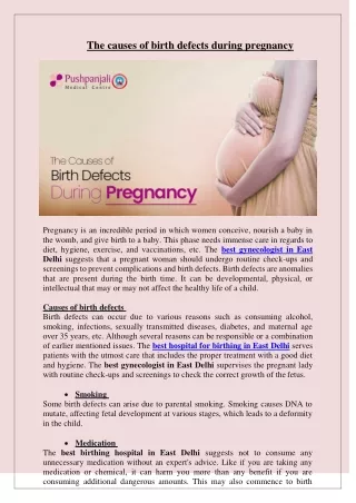
Birth injuries may result from : • Inappropriate or deficient medical skill or attention. • They may occur, despite skilled and competent obstetric care.
Incidence • as been estimated at 2-7/1,000 live births • 5-8/100,000 infants die of birth trauma • 25/100,000 die of anoxic injuries • Such injuries represent 2-3% of infant deaths.
Predisposing factors: • Macrosomia, • Prematurity, • Cephalopelvic disproportion, • Dystocia, • Prolonged or rapid delivery • Breech presentation.
Primigravida Cephalopelvic disproportion Small maternal stature maternal pelvic anomalies Prolonged or rapid labor Arrest of descent of presenting fetal part Oligohydramnios Resuscitation with CPR Abnormal presentation (breech/face) Use of forceps or vacuum extraction Versions VLBW infant or extreme prematurity Macrosomia Large fetal head Fetal anomalies Fetal neuromuscular disease Predisposing factors
Trauma during birth includes: • trauma to skin and superficial tissues • muscle trauma • nerve trauma • fractures
Birth Injuries • Soft Tissue Injuries (Abrasions, Bruising, Fat Necrosis, Lacerations) • Extracranial Bleeding (Caput succedaneum, Cephalhematoma, Subgaleal Hematoma) • Intracranial Bleeding (Subarachnoid, Epidural, Subdural, Cerebral, Cerebellar) • Nerve Injuries (Facial and Cervical Nerve Roots, Horner Syndrome, Recurrent Laryngeal Nerve)
Fractures (Clavicle, Humerus, Femur, Skull) • Dislocations • Torticollis (Sternocleidomastoid injury) • Eye Injuries (Subconjunctival and Retinal Hemorrhage) • Solid Organ Injury (liver, spleen, kidney, adrenal glands)
Trauma to skin All other skin injuries should be detected during the midwife's detailed examination of the baby immediately after birth. All trauma should be made known to the parents and reported to the baby's doctor.
Soft Tissue Injuries • Bruises and Petechiae • Can be seen in the GU area in breech presentations • Can be seen around the head and neck when there is a nuchal cord or precipitous delivery • Appearance of new bruises or petechiae after delivery warrants further investigation to r/o sepsis/DIC or bleeding disorder
Abrasions and lacerations Abrasions and lacerations should be kept clean and dry. If there are signs of infection, further medical consultation should be sought by the midwife or parents. Antibiotics may be required. Deeper lacerations may require closure with butterfly strips or sutures. Healing is usually rapid with no residual scarring
Fat Necrosis Well-circumscribed firm nodule with purplish discoloration Usually occurs after forceps use, but can occur at other sites of trauma Resolves spontaneously over weeks to months
Soft tissue trauma involves oedematous swellings and/or bruising. During labour the fetal part overlying the cervical os may be subjected to pressure, a ‘girdle of contact’, with reduced venous return and resultant congestion and oedema.
Caput succedaneum • With cephalic presentation • may be a diffuse oedematous swelling under the scalp and above the periosteum • Soft tissue swelling / edema / petechiae / ecchymoses • Crosses suture lines
A ‘false’ caput succedaneum can also occur if a vacuum extractor cup is used; because of its distinctive shape, the swelling is known as a ‘chignon • A caput succedaneum is present at birth, does not usually enlarge, can ‘pit’ on pressure • The swelling is usually self-limiting, resolving by 36 hrs of life • An abraded chignon usually heals rapidly if the area is kept clean, dry and is not irritated
Uncomplicated oedema and bruising usually resolve within days • serious complications require specific treatment, and take longer to resolve • These complications include excessive haemolysis resulting in hyperbilirubinaemia; excessive blood loss resulting in hypovolaemia, shock, anaemia and disseminated intravascular coagulation
Muscle trauma • Injuries to muscle result from tearing or when the blood supply is disrupted. • If the techniques for assisting at these stages of birth are correctly applied, torticollis is preventable
Torticollis • Lateral tilt of the neck and head typically due to a tight sternocleidomastoidmuscle • Head and neck tilt toward the involved side and chin is turned away from the involved side • The muscle length is shortened, therefore the neck is twisted to the affected side, a torticollis or wry neck
The right and left sternomastoid muscles run from the respective side of the top of the sternum, along the right or left side of the neck and are inserted into the mastoid process of the right or left temporal bone • A 1–3 cm, apparently painless, hard lump of blood and fibrous tissue is felt on the affected sternomastoid muscle • The swelling will usually resolve over several weeks to months with minimal sequelae • Stretching exercises are successful in 90% of the cases under the guidance of a physiotherapist • Surgical correction may be considered in resistant cases after 1 year of age
Nerve trauma • The nerves most commonly traumatized are the facial and brachial plexus nerves. Spinal cord injury is very rare
Facial palsy • can be caused by pressure on the facial nerves • Due to Compression may occur in the uterus but is more likely during birth by • the maternal sacral promontory or • a mis-applied forceps blade. • affected side of the face droops and the infant is unable to close the eye tightly on that side. Whencrying the mouth is pulled across to the normal side. • Spontaneous resolution is usually within 7–10 days; this may extend to months or years if the damage is severe
There is no specific treatment. If the eyelid remains open, regular instillation of eye drops lubricate the eyeball. • Feeding difficulties are usually overcome by the baby's own adaptation, although alternative feeding positions may help.
Nerve roots exiting from the spine at the fifth to eighth cervical and the first thoracic vertebrae form a matrix of nerves in the neck and shoulder; the brachial plexus. • Brachial plexus trauma usually results from excessive lateral flexion, rotation or traction of the head and neck during vaginal breech birth or when shoulder dystocia occurs. • The possible damage ranges from oedema to haemorrhage to tearing of the nerves. • There are three main injuries: Erb's palsy, Klumpke's palsy and total brachial plexus palsy. • These injuries can be unilateral or bilater
Three forms - depending on site and extent of trauma • 5th and 6th cervical roots - upper arm (Erb-Duchenne) = most common form • 8th cervical and 1st thoracic roots - lower arm (Klumpke) = extremely rare • Paralysis of entire arm = rare
Erb's palsy • There is paralysis of the shoulder and arm (not the hand) due to damage to the upper brachial plexus involving the fifth and sixth cervical nerve roots • The baby's affected arm is inwardly rotated, the elbow extended, the wrist pronated and flexed and the hand partially closed; the ‘waiter's tip position. • PlMoro, biceps, and radial reflexes are absent on the affected side. Grasp reflex is usually man’s
Klumpke's palsy • It is less common • This is due to damage to the lower brachial plexus involving the seventh and eighth cervical and the first thoracic nerve roots. • The shoulder and upper arm are unaffected but the lower arm, wrist and hand are paralysed resulting in weakness of the intrinsic muscles of the hand wrist drop and grasp reflex is absent. • Frequently with ipsilateral Horner’s syndrome (enophthalmus, ptosis, miosis, anhidrosis) - cervical sympathetic fibers from 1st thoracic root affected
Total brachial plexus palsy occur in : • Macrosomic infants and when lateral traction is exerted on the head and neck during delivery of the shoulder in a vertex presentation, • When the arms are extended over the head in a breech presentation, or • When excessive traction is placed on the shoulders.
Total brachial plexus palsy • There is complete paralysis of the shoulder, arm and hand, lack of sensation, and circulatory problems due to damage to all brachial plexus nerve roots. • If there is bilateral paralysis, spinal injury should be suspected. • All types of brachial plexus trauma will require further investigations such as X-ray and ultrasound scanning (USS) of the clavicle, arm, chest and cervical spine, and assessment of the joints. • Passive movements of the joints and limb can be initiated under the direction of a physiotherapist. • At 1 month of age, magnetic resonance imaging (MRI)can offer specific data on nerve damage.
Complete recovery within 6 months is expected for 52%of babies, 2% will have no recovery and 46% will have incomplete recovery • Babies with no functional recovery by 6 months may require microsurgical nerve repair
Phrenic nerve palsy • Lateral hyperextension of neck causes overstretching or avulsion of 3rd, 4th and 5th cervical roots which supply phrenic nerve • Respiratory distress, risk of infection, elevated hemidiaphragm, paradoxical diaphragmatic movement, atelectatic areas • If no improvement within one month, recovery unlikely and plication needs to be considered
Laryngeal nerve injury • Laryngeal nerve is part of vagus nerve - in neck behind jugular vein and carotid artery • 10% of vocal cord paralysis caused by birth trauma • Hoarse cry, stridor • Risk for aspiration, feeding problems • Recovery often over 4 - 6 weeks, sometimes longer