Download
1 / 85
880 likes | 1.68k Views
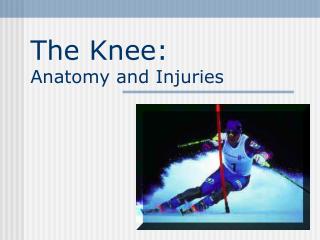
Chapter 20: The Knee and Related Structures. Complex joint that endures great amounts of trauma due to extreme amounts of stress that are regularly applied Hinge joint w/ a rotational component Stability is due primarily to ligaments, joint capsule and muscles surrounding the joint

E N D
Complex joint that endures great amounts of trauma due to extreme amounts of stress that are regularly applied • Hinge joint w/ a rotational component • Stability is due primarily to ligaments, joint capsule and muscles surrounding the joint • Designed for stability w/ weight bearing and mobility in locomotion
Knee and Related Structures • Anatomy of the Knee A. Bones i. Femur • Medial Condyle • Lateral Condyle • Medial Epicondyle • Lateral Epicondyle ii. Tibia • Tibial Plateau • Tibial Tuberosity • Gerdy’s Tubercle • Intercondylar Eminence iii. Fibula • Head iv. Patella • Largest seasmoid bone • Located within the tendon of the quadriceps femoris
Knee and Related Structures • Articulations • Femur and tibia • Femur and patella • Femur and fibula • Tibia and fibula • Menisci • Two oval fibrocartilages that deepen the articular facets of the tibia • Cushion • Maintain spacing between femur and tibia
Knee and Related Structures • Maintain stability • Medial Meniscus • “C” shaped • Lateral Meniscus • “O” shaped • Blood supply • Red-red zone = peripheral 1/3 edge = good blood supply • Red-white zone = middle 1/3 edge = minimal blood supply • White-white zone = inner 1/3 edge = avascular = no blood
Knee and Related Structures • Ligaments • Anterior Cruciate Ligament (ACL) • Anterior medial tibia to Posterior lateral femur • Prevents femur from moving posterior during wt bearing • Stabilizes tibial internal rotation • Main knee ligament stabilizer • Posterior Cruciate Ligament (PCL) • Posterior lateral tibia to Anterior medial femur • Prevents hyperextension • Prevents femur from moving anterior during wt bering
Knee and Related Structures • Medial Collateral Ligament (MCL) • Medial femoral epicondyle to Medial tibial epicondyle • Prevent valgus and external rotation forces • Has attachment to the medial meniscus • Lateral Collateral Ligament (LCL) 1. Lateral epicondyle of femur to Head of fibula
Knee and Related Structures • Joint Capsule Components • Bursa – Synovial fluid filled pouches • Reduce friction • Two dozen in and around the knee • Suprapatellar • Prepatellar • Infrapatellar • Deep infrapatellar • Fat pad • Cushions front of the knee • Separtates patellar tendon from joint capsule
Knee and Related Structures • Musculature • Knee flexion – hamstring group • Biceps femoris • Semitendinosus • Semimembranosus • Gracilis • Sartorius • Gastrocnemius • Popliteus • plantaris
Knee and Related Structures • Knee Extension – Quadriceps Group • Vastus Medialis • Vastus Lateralis • Vastus Intermedius • Rectus Femoris
Knee and Related Structures • Nerve Supply • Tibial nerve = hamstring and gastrocnemius • Common peroneal nerve = proximal fibula head = contusion causes sensory and motor deficits distally • Femoral nerve • Blood Supply i. Popliteal artery = stem of femoral artery
Knee and Related Structures • Leg Alignment Deviations • Predispose to injury • Patellar malalignment • Genu valgum • Genu varum • Genu recurvatum • Leg-Length and Patella Discrepancies • Anatomical leg length (true leg length) • ASIS to Lateral Malleolus • Anatomical leg length (functional leg length) • Umbilicus to Medial Malleolus • Girth Measurement • Q-Angle Measurement 1. ASIS to Mid-patella to Tibial Tuberosity
Knee and Related Structures • Special Tests for Knee Joint Stability • Valgus Stress Test • Varus Stress Test • Anterior Drawer • Lachman Drawer Test • Pivot Shift Test • Posterior Drawer Test • Posterior Sag Test • McMurray’s Test • Apley Compression Test • Apley Distraction Test • Patellar Compression, Grinding, Apprehension, Chandelier Tests
Functional Anatomy • Movement of the knee requires flexion, extension, rotation and the arthrokinematic motions of rolling and gliding • Rotational component involves the “screw home mechanism” • As the knee extends it externally rotates because the medial femoral condyle is larger than the lateral • Provides increased stability to the knee • Popliteus “unlocks” knee allowing knee to flex
Capsular ligaments are taut during full extension and relaxed w/ flexion • Allows rotation to occur • Deeper capsular ligaments remain taut to keep rotation in check • PCL prevents excessive internal rotation, guides the knee in flexion, and acts as drag during initial glide phase of flexion • ACL stops excessive internal rotation, stabilizes the knee in full extension and prevents hyperextension
Range of motion includes 140 degrees of motion • Limited by shortened position of hamstrings, bulk of hamstrings and extensibility of quads • Patella aids knee during extension, providing a mechanical advantage • Distributes compressive stress on the femur by increasing contact between patellar tendon and femur • Protects patellar tendon against friction • When moving from extension to flexion the patella glides laterally and further into trochlear groove
Kinetic Chain • Directly affected by motions and forces occurring at the foot, ankle, lower leg, thigh, hip, pelvis, and spine • With the kinetic chain forces must be absorbed and distributed • If body is unable to manage forces, breakdown to the system occurs • Knee is very susceptible to injury resulting from absorption of forces
Assessing the Knee Joint • Determining the mechanism of injury is critical • History- Current Injury • Past history • Mechanism- what position was your body in? • Did the knee collapse? • Did you hear or feel anything? • Could you move your knee immediately after injury or was it locked? • Did swelling occur? • Where was the pain
History - Recurrent or Chronic Injury • What is your major complaint? • When did you first notice the condition? • Is there recurrent swelling? • Does the knee lock or catch? • Is there severe pain? • Grinding or grating? • Does it ever feel like giving way? • What does it feel like when ascending and descending stairs? • What past treatment have you undergone?
Observation • Walking, half squatting, going up and down stairs • Swelling, ecchymosis, • Leg alignment • Genu valgum and genu varum • Hyperextension and hyperflexion • Patella alta and baja • Patella rotated inward or outward • May cause a combination of problems • Tibial torsion, femoral anteversion and retroversion
Tibial torsion • An angle that measures less than 15 degrees is an indication of tibial torsion • Femoral Anteversion and Retroversion • Total rotation of the hip equals ~100 degrees • If the hip rotates >70 degrees internally, anteversion of the hip may exist • INSERT 20-9
Knee Symmetry or Asymmetry • Do the knees look symmetrical? Is there obvious swelling? Atrophy? • Leg Length Discrepancy • Anatomical or functional • Anatomical differences can potentially cause problems in all weight bearing joints • Functional differences can be caused by pelvic rotations or mal-alignment of the spine
Medial tibial plateau Medial femoral condyle Adductor tubercle Gerdy’s tubercle Lateral tibial plateau Lateral femoral condyle Lateral epicondyle Head of fibula Tibial tuberosity Superior and inferior patella borders (base and apex) Around the periphery of the knee relaxed, in full flexion and extension Palpation - Bony
Vastus medialis Vastus lateralis Vastus intermedius Rectus femoris Quadriceps and patellar tendon Sartorius Medial patellar plica Anterior joint capsule Iliotibial Band Arcuate complex Medial and lateral collateral ligaments Pes anserine Medial/lateral joint capsule Semitendinosus Semimembranosus Gastrocnemius Popliteus Biceps Femoris Palpation - Soft Tissue
Palpation of Swelling • Intra vs. extracapsular swelling • Intracapsular may be referred to as joint effusion • Swelling w/in the joint that is caused by synovial fluid and blood is a hemarthrosis • Sweep maneuver • Ballotable patella - sign of joint effusion • Extracapsular swelling tends to localize over the injured structure • May ultimately migrate down to foot and ankle
Special Tests for Knee Instability • Use endpoint feel to determine stability • MRI may also be necessary for assessment • Classification of Joint Instability • Knee laxity includes both straight and rotary instability • Translation (tibial translation) refers to the glide of tibial plateau relative to the femoral condyles • As the damage to stabilization structures increases, laxity and translation also increase • Valgus and Varus Stress Tests • Used to assess the integrity of the MCL and LCL respectively • Testing at 0 degrees incorporates capsular testing while testing at 30 degrees of flexion isolates the ligaments
Anterior Cruciate Ligament Tests • Drawer test at 90 degrees of flexion • Tibia sliding forward from under the femur is considered a positive sign (ACL) • Should be performed w/ knee internally and externally to test integrity of joint capsule
Lachman Drawer Test • Will not force knee into painful flexion immediately after injury • Reduces hamstring involvement • At 30 degrees of flexion an attempt is made to translate the tibia anteriorly on the femur • A positive test indicates damage to the ACL
Pivot Shift Test • Used to determine anterolateral rotary instability • Position starts w/ knee extended and leg internally rotated • The thigh and knee are then flexed w/ a valgus stress applied to the knee • Reduction of the tibial plateau (producing a clunk) is a positive sign
Posterior Cruciate Ligament Tests • Posterior Drawer Test • Knee is flexed at 90 degrees and a posterior force is applied to determine translation posteriorly • Positive sign indicates a PCL deficient knee
Posterior Sag Test (Godfrey’s test) • Athlete is supine w/ both knees flexed to 90 degrees • Lateral observation is required to determine extent of posterior sag while comparing bilaterally
Instrument Assessment of the Cruciate Ligaments • A number of devices are available to quantify AP displacement of the knee • KT-2000 arthrometer, Stryker knee laxity tester and Genucom can be used to assess the knee • Test can be taken pre & post-operatively and through rehab
Meniscal Tests • McMurray’s Meniscal Test • Used to determine displaceable meniscal tear • Leg is moved into flexion and extension while knee is internally and externally rotated in conjunction w/ valgus and varus stressing • A positive test is found w/ clicking and popping response
Apley’s Compression Test • Hard downward pressure is applied w/ rotation • Pain indicates a meniscal injury • Apley’s Distraction Test • Traction is applied w/ rotation • Pain will occur if there is damage to the capsule or ligaments • No pain will occur if it is meniscal
Girth Measurements • Changes in girth can occur due to atrophy, swelling and conditioning • Must use circumferential measures to determine deficits and gains during the rehabilitation process • Measurements should be taken at the joint line, the level of the tibial tubercle, belly of the gastrocnemius, 2 cm above the superior border of the patella, and 8-10 cm above the joint line • Subjective Rating • Used to determine patient’s perception of pain, stability and functional performance
Functional Examination • Must assess walking, running, turning and cutting • Co-contraction test, vertical jump, single leg hop tests and the duck walk • Resistive strength testing • Q-Angle • Lines which bisects the patella relative to the ASIS and the tibial tubercle • Normal angle is 10 degrees for males and 15 degrees for females • Elevated angles often lead to pathological conditions associated w/ improper patella tracking
Palpation of the Patella • Must palpate around and under patella to determine points of pain • Patella Grinding, Compression and Apprehension Tests • A series of glides and compressions are performed w/ the patella to determine integrity of patellar cartilage
Prevention of Knee Injuries • Physical Conditioning and Rehabilitation • Total body conditioning is required • Strength, flexibility, cardiovascular and muscular endurance, agility, speed and balance • Muscles around joint must be conditioned (flexibility and strength) to maximize stability • Must avoid abnormal muscle action through flexibility • In an effort to prevent injury, extensibility of hamstrings, erector spinae, groin, quadriceps and gastrocnemius is important