Download
1 / 14
140 likes | 225 Views
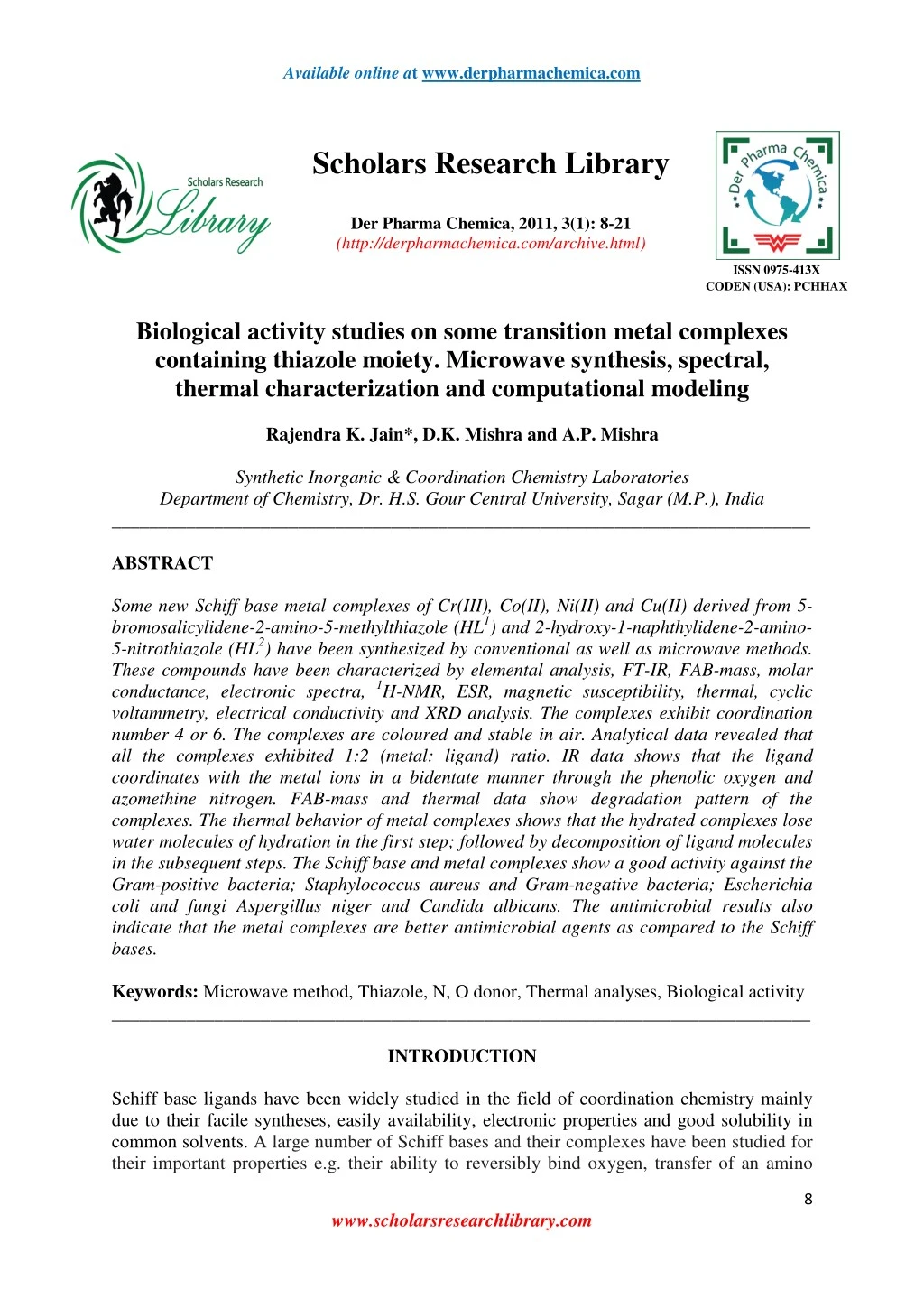
Some new Schiff base metal complexes of Cr(III), Co(II), Ni(II) and Cu(II) derived from 5-<br>bromosalicylidene-2-amino-5-methylthiazole (HL1<br>) and 2-hydroxy-1-naphthylidene-2-amino5-nitrothiazole<br>(HL2<br>) have been synthesized by conventional as well as microwave methods.<br>These compounds have been characterized by elemental analysis, FT-IR, FAB-mass, molar<br>conductance, electronic spectra, 1H-NMR, ESR, magnetic susceptibility, thermal, cyclic<br>voltammetry, electrical conductivity and XRD analysis. The complexes exhibit coordination<br>number 4 or 6. The complexes are coloured and stable in air. Analytical data revealed that<br>all the complexes exhibited 1:2 (metal: ligand) ratio. IR data shows that the ligand<br>coordinates with the metal ions in a bidentate manner through the phenolic oxygen and<br>azomethine nitrogen. FAB-mass and thermal data show degradation pattern of the<br>complexes. The thermal behavior of metal complexes shows that the hydrated complexes lose<br>water molecules of hydration in the first step; followed by decomposition of ligand molecules<br>in the subsequent steps. The Schiff base and metal complexes show a good activity against the<br>Gram-positive bacteria; Staphylococcus aureus and Gram-negative bacteria; Escherichia<br>coli and fungi Aspergillus niger and Candida albicans. The antimicrobial results also indicate that the metal complexes are better antimicrobial agents as compared to the Schiff bases.

E N D
Available online at www.derpharmachemica.com Scholars Research Library Der Pharma Chemica, 2011, 3(1): 8-21 (http://derpharmachemica.com/archive.html) ISSN 0975-413X CODEN (USA): PCHHAX Biological activity studies on some transition metal complexes containing thiazole moiety. Microwave synthesis, spectral, thermal characterization and computational modeling Rajendra K. Jain*, D.K. Mishra and A.P. Mishra Synthetic Inorganic & Coordination Chemistry Laboratories Department of Chemistry, Dr. H.S. Gour Central University, Sagar (M.P.), India ___________________________________________________________________________ ABSTRACT Some new Schiff base metal complexes of Cr(III), Co(II), Ni(II) and Cu(II) derived from 5- bromosalicylidene-2-amino-5-methylthiazole (HL1) and 2-hydroxy-1-naphthylidene-2-amino- 5-nitrothiazole (HL2) have been synthesized by conventional as well as microwave methods. These compounds have been characterized by elemental analysis, FT-IR, FAB-mass, molar conductance, electronic spectra, 1H-NMR, ESR, magnetic susceptibility, thermal, cyclic voltammetry, electrical conductivity and XRD analysis. The complexes exhibit coordination number 4 or 6. The complexes are coloured and stable in air. Analytical data revealed that all the complexes exhibited 1:2 (metal: ligand) ratio. IR data shows that the ligand coordinates with the metal ions in a bidentate manner through the phenolic oxygen and azomethine nitrogen. FAB-mass and thermal data show degradation pattern of the complexes. The thermal behavior of metal complexes shows that the hydrated complexes lose water molecules of hydration in the first step; followed by decomposition of ligand molecules in the subsequent steps. The Schiff base and metal complexes show a good activity against the Gram-positive bacteria; Staphylococcus aureus and Gram-negative bacteria; Escherichia coli and fungi Aspergillus niger and Candida albicans. The antimicrobial results also indicate that the metal complexes are better antimicrobial agents as compared to the Schiff bases. Keywords: Microwave method, Thiazole, N, O donor, Thermal analyses, Biological activity ___________________________________________________________________________ INTRODUCTION Schiff base ligands have been widely studied in the field of coordination chemistry mainly due to their facile syntheses, easily availability, electronic properties and good solubility in common solvents. A large number of Schiff bases and their complexes have been studied for their important properties e.g. their ability to reversibly bind oxygen, transfer of an amino 8 www.scholarsresearchlibrary.com
Rajendra K. Jain et al __________________________________________________________________________ Der Pharma Chemica, 2011, 3 (1): 8-21 group and complexing ability towards some toxic metals [1-3]. The high affinity for the chelation of the Schiff bases towards the transition metal ions is utilized in preparing their solid complexes. Schiff base metal complexes have been useful to design and develop some models for biological systems. Transition metal complexes which usually contain nitrogen, sulphur/or oxygen as ligand atoms are becoming increasingly important because these Schiff base can bind with different metal centers involving various coordination sites and allow successful synthesis of metallic complexes with interesting stereochemistry [4-7]. Heterocyclic compounds are widely distributed in the nature and essential to many biochemicals, analytical and industrial processes. Compounds containing these heterocycles have important properties in the field of material science and biological systems. Various heterocycles especially 2-aminothiazoles are a remarkably versatile group of compounds that have found recent applications in the drug development and production of dyes. Thiazole derivatives play important role in many biochemical reactions. The molecular systems having chelating molecular designing and incorporating thiazole ring have been examined in the field of mycology for the development of metal based drugs. In several decades, efforts have been made to design and development of these drugs with thiazole based molecular devices. The basic characteristics of these metal based drugs for their drug activity is their penetration power through pathogenic cell membrane and their affinity for the genetic material (DNA and RNA) of the pathogens. Schiff base from 2-hydroxy-1-naphthaldehyde have often been used as chelating ligand in the field of coordination chemistry [8-10]. Microwave-assisted synthesis is a branch of green chemistry. Microwave-assisted synthesis has gained much attention in recent years. Microwave-assisted organic synthesis is characterized by the spectacular accelerations produced in many reactions as a consequence of the heating rate, which cannot be reproduced by thermal heating. Higher yields, milder reaction conditions, shorter reaction times can be used and many processes can be improved. The applications of microwave irradiation are used for carrying out chemical transformations, which are pollution free and eco-friendly. The basis of this technique of synthesis is much faster with higher yields compared to conventional heating. Reports on the synthesis of metal complexes by microwave methods have been comparatively less [11-16]. In the present paper, we have described the coordination behavior of novel Schiff bases (Fig. 1) derived from the condensation of 5-bromosalicylaldehyde with 2-amino-5-methylthiazole (HL1) and 2-hdroxy-1-naphthaldehyde with 2-amino-5-nitrothiazole (HL2) towards some transition elements, which may help in more understanding of the mode of chelation of ligands towards metals. For this purpose the complexes of Cr(III), Co(II), Ni(II) and Cu(II) ions with HL1 and HL2 have been synthesized by both conventional as well as microwave methods and characterized by the various physicochemical and spectral analyses. These ligands coordinate with metal ions in a bidentate manner through the phenolic oxygen and azomethine nitrogen. MATERIALS AND METHODS Experimental Chemical materials and instruments All the used chemicals and solvents were of Anal R grade. All the reagents used for the preparation of the Schiff bases were obtained from Sigma Aldrich. Metal salts were purchased from Loba Chemie. Elemental analyses were performed on an Elementar Vario EL III Carlo Erba 1108 analyzer. FAB-mass spectra were recorded on a JEOL SX 102/DA 6000 Mass Spectrometer using argon/xenon (6 kV, 10 mA) as the FAB gas. The accelerating 9 www.scholarsresearchlibrary.com
Rajendra K. Jain et al __________________________________________________________________________ Der Pharma Chemica, 2011, 3 (1): 8-21 voltage was 10 kV and the spectra were recorded at room temperature. Electronic spectra (in DMSO) were recorded on Perkin Elmer Lambda-2B-spectrophotometer. Molar conductance measurements were conducted using 10-3 M solutions of the complexes in DMSO on Elico- CM 82 Conductivity Bridge at room temperature. Magnetic susceptibility measurements were carried out on a Gouy balance at room temperature using mercuric tetrathiocyanato cobaltate(II) as the calibrant. Diamagnetic corrections were applied in compliance with Pascal’s constant. FT-IR spectra were recorded in KBr medium on a Perkin Elmer RX1 spectrophotometer in wave number region 4000-400 cm-1. 1H NMR spectra were recorded on a JEOL AL300 FTNMR spectrometer employing TMS as internal reference and DMSO-d6 as solvent. Cyclic voltammetry was carried out with a BAS-100 Epsilon electrochemical analyzer having an electrochemical cell with a three electrode systems. Ag/AgCl was used as a reference electrode, glassy carbon as working electrode and platinum wire as an auxiliary. 0.1 M NaClO4 was used as supporting electrolyte and DMSO as solvent. All measurements were performed at room temperature under a nitrogen atmosphere. X-band EPR spectra were recorded on a Varian E-112 spectrometer at room temperature operating at the X-band region with 100 kHz modulation frequency, 5 mw microwave power and 1 G modulation amplitude using TCNE as the internal standard. Thermogravimetric analysis was carried out under atmospheric condition with a heating rate 10 °C min-1 on TGA Q500 universal V4.5A TA instrument. Microwave assisted synthesis were carried out in open glass vessel on a modified microwave oven model 2001 ETB with rotating tray and a power source 230 V, microwave energy output 800 W and microwave frequency 2450 MHz. A thermocouple device was used to monitor the temperature inside the vessel of the microwave. The microwave reactions were performed using on/off cycling to control the temperature. Biological activity The in vitro biological activity of the investigated Schiff base and its metal complexes was tested against the bacteria Escherichia coli and Staphylococcus aureus by disc diffusion method using nutrient agar as medium and streptomycin as control. The antifungal activities of the compounds were also tested by the Well diffusion method against the fungi Aspergillus niger and Candida albicans, on potato dextrose agar as the medium and miconazole as control. The stock solution was prepared by dissolving the compounds in DMSO. In a typical procedure, a well was made on agar medium inoculated with microorganism. The well was filled with the test solution using a micropipette and the plate was incubated 24 h for bacteria at 37 °C and 72 h for fungi at 30 °C. During this period, the test solution diffused and the growth of the inoculated microorganism was affected. The inhibition zone was developed, at which the concentration was noted. Conventional method for the Synthesis of Schiff bases HL1 Schiff base was synthesized by the condensation of equimolar ratio of 5- bromosalicylaldehyde (0.201 g, 10 mmol) with 2-amino-5-methylthiazole (0.114 g, 10 mmol) dissolved in ethanol. The resulting reaction mixture was stirred well, refluxed for 3-4 h and then allowed to cool overnight. The coloured solid precipitate of Schiff base obtained was filtered, washed with cold ethanol several times and dried in air at room temperature and finally stored under reduced pressure in a CaCl2 desiccator. The purity of synthesized compounds was checked by TLC using silica gel G (yield: 80%). Characteristic 1H NMR peaks in HL1 are (DMSO-d6, TMS, δ ppm), 8.906 (s,-CH=N), 12.312 (s, -OH), 2.492 (s, - CH3), 7.405-7.765 (m, aromatic proton), 6.926 (s, thiazole proton). FAB-mass (m/z) calc.: 297, found: 299. 10 www.scholarsresearchlibrary.com
Rajendra K. Jain et al __________________________________________________________________________ Der Pharma Chemica, 2011, 3 (1): 8-21 HL2 Schiff base was synthesized by the condensation of 1:1 ratio of 2-hydroxy-1- naphthaldehyde (0.172 g, 10 mmol) with 2-amino-5-nitrothiazole (0.145 g, 10 mmol) dissolved in ethanol. The resulting reaction mixture was refluxed for 3-4 h and then allowed to cool overnight. The coloured solid precipitate of Schiff base obtained was filtered, washed with cold ethanol, finally recrystallized from ethanol, ether and dried in air at room temperature, preserved in a CaCl2 desiccator. The purity of synthesized compounds was checked by TLC using silica gel G (yield: 77%). Characteristic 1H NMR peaks in HL2 are (DMSO, TMS, δ ppm), 8.985 (s,-CH=N), 12.468 (s, -OH), 7.345-7.709 (m, aromatic proton), 7.105 (s, thiazole proton). FAB-mass (m/z) calc.: 299, found: 300. Microwave method for the Synthesis of Schiff bases The equimolar (1:1) ratio of 5-bromosalicylaldehyde (0.201 g, 10 mmol) with 2-amino-5- methylthiazole (0.114 g, 10 mmol) and 2-hydroxy-1-naphthaldehyde (0.172 g, 10 mmol) with 2-amino-5-nitrothiazole (0.145 g, 10 mmol) were mixed thoroughly in a grinder. The reaction mixture was then irradiated by the microwave oven by taking 3-4 ml solvent. The reaction was completed in a short time (4-5 min) with higher yields. The resulting product was then recrystallized with ethanol, finally dried under reduced pressure over anhydrous CaCl2 in a desiccator. The progress of the reaction, purity of the product was monitored by TLC using silica gel G (yield: 88-90%). Conventional method for the Synthesis of metal complexes The metal complexes were prepared by the mixing of (50 ml) ethanolic solution of CrCl3.6H2O/CoCl2.6H2O/NiCl2.6H2O/CuCl2.2H2O with the (50 ml) ethanolic solution of Schiff bases (HL1/HL2) in 1:2 (metal:ligand) ratio. The resulting mixture was refluxed on water bath for 5-9 h. A coloured product appeared on standing and cooling the above solution. The precipitated complex was, filtered washed with ether and recrystallized with ethanol several times and dried under the reduced pressure over anhydrous CaCl2 in a desiccator. It was further dried in electric oven at 50-70 °C (yield: 58-74%). Microwave method for the Synthesis of metal complexes The ligand and the metal salts were mixed in 1:2 (metal:ligand) ratio in a grinder. The reaction mixture was then irradiated by the microwave oven by taking 3-4 ml solvent. The reaction was completed in a short time (6-10 min) with higher yields. The resulting product was then recrystallized with ethanol and ether and finally dried under reduced pressure over anhydrous CaCl2 in a desiccator. The progress of the reaction and purity of the product was monitored by TLC using silica gel G (yield: 75-86%). RESULTS AND DISCUSSION As a result of microwave-assisted synthesis, it was observed that the reaction was completed in a short time with higher yields compared to the conventional method. In the microwave method homogeneity of reaction mixture was increased by the rotating of reaction platform tray. The confirming of the results was also checked by the repeating of the synthesis process. Comparative study results obtained by microwave assisted synthesis; versus conventional heating method is that some reactions which required 3.1-8.6 h. by conventional method, was completed within 4.2-8.9 min. by the microwave irradiation technique, yields have been improved from 58-80% to 81-90%. All the metal complexes are coloured, solid and stable towards air and moisture at room temperature. They decompose on heating at high temperature, more or less soluble in 11 www.scholarsresearchlibrary.com
Rajendra K. Jain et al __________________________________________________________________________ Der Pharma Chemica, 2011, 3 (1): 8-21 common organic solvents. The comparative results of conventional and microwave methods and analytical data of the compounds, together with their physical properties are consistent with proposed molecular formula are given in Table 1. All the metal chelates have 1:2 (metal:ligand) stoichiometry. The observed molar conductance of the complexes in DMSO at room temperature are consistent with the non-electrolytic nature of the complexes except Cr(III) complex of both the Schiff base ligands, which it was electrolytic nature. FAB-mass spectrum The FAB-mass spectra suggested that all the complexes have a monomeric nature. These complexes show molecular ion peaks in good agreement with the empirical formula suggested by elemental analyses. The FAB-mass spectrum of the [Co(L1)2(H2O)2] complex shows a characteristic molecular ion (M+) peak at m/z = 688, which corresponds to the molecular weight of the complex for a monomeric structure. The mass spectrum shows multiple peaks representing successive degradation of the complex molecule by the formation of different fragments (Scheme-1). The peaks of appreciable intensity have been observed at m/z values, obs. (cal.) – 688(687), 654(651), 619(651), 467(461), 290(295), 117(119) suggesting the fragmentation pattern. The m/z value 688 corresponds to nearest composition of the [Co(L1)2(H2O)2] and 117 corresponds to Co metal with chelated O, N ligand moiety. The FAB-mass of the [Cr(L2)2(H2O)2]Cl complex exhibited the molecular ion (M+) peak at m/z = 722 suggesting the monomeric nature of the complex. The other important peaks of appreciable intensity have been observed at m/z values, obs. (cal.) – 680(684), 651(648), 560 (556), 391(390), 107(112) suggesting the ion species after the successive fragmentation of different groups. The intensities of these peaks give the idea of the stabilities of the fragments. The m/z value 722 corresponds to nearest composition [Cr(L2)2(H2O)2]Cl and 107 to Cr metal with chelated N, O ligand moiety [17-18]. IR spectra The data of the IR spectra of Schiff base ligand, its metal complexes are listed in Table 2. The IR spectra of the complexes were compared with those of the free ligand in order to determine the involvement of coordination sites in chelation. Characteristic peaks in the spectra of the ligand and complexes were considered and compared. IR spectrum of the HL1 ligand exhibit the most characteristic bands at 3298 cm-1ν(O-H), 1628 cm-1ν(C=N, azomethine), 1536 cm-1ν(C=N, thiazole), 1244 cm-1ν(C-O) and 727 cm-1 ν(C-S-C). The metal complexes of Cr(III), Co(II) and Ni(II) show a broad band at (3362- 3381cm-1) which may be due to νstr(OH). A medium intensity band at 813 cm-1, 808 cm-1 and 815 cm-1 in Cr(III), Co(II) and Ni(II) respectively, suggests the presence of coordinated water in these complexes. But these bands are not observed in Cu(II) complex indicate the absence of water molecules in the Cu(II) complex. The band 1628 cm-1 due to the azomethine group of the Schiff base has shifted to lower frequency (1596-1608 cm-1) after complexation, indicating the bonding of nitrogen of the azomethine group to the metal ion, this can be explained by the donation of electrons from nitrogen to the empty d-orbital of the metal atom [19-20]. The phenolic C-O stretching vibration that appeared at 1244 cm-1 in Schiff base shift towards higher frequencies (27-35 cm-1) in the complexes. This shift confirms the participation of oxygen in the C-O-M bond. In the IR spectra of the complexes, the stretching vibration of the free ligand ν(O-H) 3298 cm-1 is not observed, suggesting deprotonation of the hydroxyl group and formation of M-O bonds. In the low frequency region, the band of weak 12 www.scholarsresearchlibrary.com
Rajendra K. Jain et al __________________________________________________________________________ Der Pharma Chemica, 2011, 3 (1): 8-21 intensity observed for the complexes in the region 524-544 cm-1 is attributed to (M-O), in the region 489-494 cm-1 to (M-N). The ν(C=N) at 1536 cm-1 and ν(C-S-C) at 727 cm-1 of the thiazole ring remain unchanged suggested that thiazole group does not coordinate to metal ion by neither nitrogen nor sulpher atom [21]. IR spectrum of the HL2 ligand show the most characteristic bands at 3308 cm-1ν(O-H), 1631 cm-1ν(C=N, azomethine), 1548 cm-1ν(C=N, thiazole), 1251 cm-1ν(C-O) and 738cm-1ν(C- S-C). All the metal complexes show a broad band at (3378-3410 cm-1) which may be due to νstr(OH). A medium intensity band at 812 cm-1, 817 cm-1 and 819 cm-1 in Cr(III), Co(II) and Ni(II) respectively, suggests the presence of coordinated water molecules in these complexes. But this band are not observed in Cu(II) complex indicate the absence of coordinated water molecules in the Cu(II) complex. The band 1631 cm-1 due to the azomethine group of the Schiff base has shifted to lower frequency (1599-1607 cm-1) after complexation. This indicates the involvement of azomethine nitrogen to the metal ion. The phenolic C-O stretching vibration that appeared at 1251 cm-1 in Schiff base shift towards higher frequencies (30-39 cm-1) in the complexes. This shift confirms the participation of oxygen in the C-O-M bond. In the IR spectra of the complexes, the stretching vibration of the free ligand ν(O-H) 3308 cm-1 is not observed, suggesting deprotonation of the hydroxyl group and formation of M-O bond. In the low frequency region, the band of weak intensity observed for the complexes in the region 537-546 cm-1 is attributed to (M-O), in the region 489-501 cm-1 to (M-N). The ν(C=N) at 1548 cm-1 and ν(C-S-C) at 738 cm-1 of the thiazole ring remain unchanged suggested that thiazole group does not participate in coordination to the metal ion [20-21]. The IR data of both the Schiff base and its metal complexes show that the Schiff bases (HL1 and HL2) is coordinated to the metal ion in bidentate manner with ON donor sites of deprotonated phenolic oxygen and azomethine nitrogen. Electronic spectra and magnetic moment The nature of the ligand field around the metal ion has been deduced from the electronic spectra. The electronic spectra of Cr(III) complex of HL1 observed bands in the region 18205 cm-1, 24650 cm-1 and 35450 cm-1,which may be assigned to 4A2g→4T2g(F) (ν1), 4A2g→4T1g(F) (ν2) and 4A2g→4T1g(P) (ν3) transitions, respectively. The magnetic moment is 3.91 B.M. Thus the octahedral geometry has been suggested for this complex. The electronic spectrum of Co(II) complex of HL1 exhibit three bands in the range of 9344 cm-1, 15352 cm-1 and 20328 cm-1 which have tentatively been assigned to 4T1g→4T2g(F) (ν1), 4T1g→4A2g(F) (ν2) and 4T1g→4T1g(P) (ν3) transitions, respectively. The value of magnetic moment is 5.09 B.M.; which indicates the presence of Co(II) complex in octahedral geometry. The absorption spectrum of Ni(II) complex of HL1 display three bands at 10568 cm-1, 18846 cm-1 and 23954 cm-1, which have tentatively been assigned to the transitions 3A2g→3T2g (F)(ν1), 3A2g→3T1g (F) (ν2) and 3A2g→3T1g (P) (ν3) respectively. The value of magnetic moment is 3.11 B.M.; therefore octahedral geometry has been suggested for this complex. The electronic spectrum of the Cu(II) complex of HL1 show two bands at 13388 cm-1 and 17660 cm-1 assignable to 2B1g→2B2g and 2B1g→2Eg transitions, respectively. Since the value 13 www.scholarsresearchlibrary.com
Rajendra K. Jain et al __________________________________________________________________________ Der Pharma Chemica, 2011, 3 (1): 8-21 of magnetic moment is found 1.98 B.M.; therefore square planar geometry has been suggested for Cu(II) complex. The Cr(III) complex of HL2 show electronic spectral bands at 17930 cm-1, 24691 cm-1 and 35872 cm-1 these are tentatively assigned to 4A2g→4T2g(F) (ν1), 4A2g→4T1(F) (ν2) and 4A2g→4T1g(P) (ν3) transitions, respectively. The magnetic moment value is 3.94 B.M. Thus the octahedral structure has been suggested for this complex. The electronic spectrum of Co(II) complex of HL2 show three bands at 9505 cm-1, 15680 cm- 1 and 19792 cm-1, these are tentatively assigned to 4T1g→4T2g(F) (ν1), 4T1g→4A2g(F) (ν2) and 4T1g→4T1g (P) (ν3) transitions, respectively. The magnetic susceptibility value is 5.12 B.M.; which is an indicative of octahedral geometry. The electronic spectrum of the Ni(II) complex of HL2 show three bands at 11120 cm-1, 18677 cm-1 and 23896 cm-1 assignable to 3A2g→3T2g (F)(ν1), 3A2g→3T1g (F)(ν2) and 3A2g→3T1g (P)(ν3) transitions, respectively. The value of magnetic moment is 3.12 B.M.; therefore octahedral geometry has been suggested for this complex. The electronic spectrum of the Cu(II) complex of HL2 show two bands at 13982 cm-1 and 19112 cm-1 assignable to 2B1g→2B2g and 2B1g→2Eg transitions, respectively. The value of magnetic moment for this complex is 2.02 B.M.; thus the square planar geometry has been suggested for Cu(II) complex [22-26]. ESR spectra The ESR spectra of Cu(II) provide information about the extent of the delocalization of unpaired electron. The X-band ESR spectra of Cu(II) complexes were recorded in the solid state at room temperature, their g║, g┴, ∆g, gav, G have been calculated. The values of ESR parameters g║, g┴, gav,∆g, G for Cu(II) complex of HL1 are 2.2298, 2.1486, 2.1757, 0.0812, 1.5539respectively. Similarly, the corresponding values for Cu(II) complex of HL2 are 2.2564, 2.1831, 2.2075, 0.0733, 1.4054 respectively. ESR spectra of the complexes revealed two g values (g║ and g┴). Since the g║ and g┴ values are closer to 2 and g║> g┴suggesting a tetragonal distortion around the Cu(II) ion. The trend g║> g┴>ge(2.0023)shows that the unpaired electron is localized in dX2-Y2 orbital in the ground state of Cu(II), spectra are characteristic of axial symmetry. The g║>2.3 is characteristic of an ionic environment and g║<2.3 indicates a covalent environment in metal ligand bonding. The g║ values for these complexes are less than 2.3 suggesting the environment is covalent [27]. The exchange coupling interaction between two Cu(II) ions is explained by Hathaway expression G = (g║-2.0023)/ (g┴-2.0023). According to Hathaway [28-29], if the value G is greater than four (G>4.0), the exchange interaction is negligible; whereas when the value of G is less than four (G<4) a considerable exchange coupling is present in solid complex. The G values for these Cu(II) complexes are less than four indicate, considerable exchange interaction in the complexes. Thermal analyses The thermal behavior of metal complexes shows that the hydrated complexes lose molecules of hydration first; followed by decomposition of ligand molecules in the subsequent steps. 14 www.scholarsresearchlibrary.com
Rajendra K. Jain et al __________________________________________________________________________ Der Pharma Chemica, 2011, 3 (1): 8-21 The thermal degradation behavior of the Ni(II) complex of HL1 has been studied by thermogravimetric analysis. The TGA curve of the complex shows no loss in weight upto 60 °C. Above this temperature a weight loss has been observed corresponding to one lattice water and two coordinated water molecules (Remaining wt.%, obs./calc., 92.60/92.34). After 225 °C, a weight loss has been observed in general upto 428 °C, indicating decomposition of non-coordinated part of the ligand (Remaining wt.%, obs./calc., 46.3/41.84). The decomposition of remaining ligand moiety occurs between 500-700 °C, above 700 °C a horizontal curve has been observed suggesting the ultimate pyrolysis product as metal oxide (Remaining wt.%, obs./calc., 23.25/21.78). The DTG curve of the complex shows peak at 150, 388, 636 °C [30-31]. The thermogram of the Co(II) complex of HL2 shows that the complex stable upto 105 °C. This indicates the absence of lattice water molecule in the complex. On increasing the temperature decomposition started between the temperature range 105-200 °C, corresponding to elimination of two coordinated water molecules (Remaining wt.%, obs./cal., 95.10/94.80). After this temperature, a loss in weight has been observed in general up to 340 °C corresponding to the loss of partially decomposed ligand part from the complex (Remaining wt.%, obs./cal., 64.32/62.61). Above 460 °C, a weight loss has been occurs upto 690 °C.This indicates the elimination of the remaining thermally degradable part of the complex. After 670 °C a horizontal curve has been observed which corresponds to a mixture of metal oxide as an ultimate pyrolysis product (Remaining wt.%, obs./cal., 24.45/21.40). DTG peak has been observed for the Co(II) complex at 174, 327, 643 °C [32]. The thermal analysis evaluating the thermal stability of the metal complexes, this study also helped to characterize the metal complexes. Electrochemical studies The electrochemical properties of Cu(II) complex of HL1 have been studied by cyclic voltametry (CV) under a nitrogen atmosphere in a DMSO solution in the potential range +1.2 to -1.0 V versus Ag/AgCl reference electrode. The Cu(II) complex of HL1 show reduction peak at Epc = -577 mV with a corresponding oxidation peak at Epa = -154 mV at a scan rate 100 mV/s. The peak separation ∆Ep = 423 mV of this couple. Thus, the cyclic voltametric studies gave evidence to a quasi-reversible Cu(II)/Cu(I) couple [33-34]. Molecular modeling Theoretical calculations have paid a considerable attention to the characterization. The optimized geometry for the Cu(II) complex of HL1 ligand by computing the minimum steric energy and the theoretical physical parameters such as, bond length and bond angles calculated by using MM2 method. The optimized structure of the complex is given in Fig. 2. Selected calculated parameters in coordination sphere are given in Table 3[35, 36]. Antimicrobial activities The in vitro Antimicrobial activity of the synthesized Schiff base ligands and their corresponding metal complexes on selected bacteria E. coli and S. aureus and two fungi A. niger and C. albicans was carried out. All of the tested compounds showed good biological activity against microorganism. On comparing the biological activity of the Schiff base and its metal complexes with the standard bactericide and fungicide, it is show that the some metal complexes have good activity as compared to the standard but all the complexes are more active than their respective ligands. 15 www.scholarsresearchlibrary.com
Rajendra K. Jain et al __________________________________________________________________________ Der Pharma Chemica, 2011, 3 (1): 8-21 Table 1.The comparative results of conventional and microwave methods, analytical and physical data of the compounds Reaction period CM (MM) 3.1 (4.2) 8.0 (8.2) 7.3 (6.9) 6.5 (7.0) 6.1 (7.1) 3.9 (5.1) 8.6 (8.9) 7.0 (7.8) 6.8 (8.1) 7.6 (7.2) Yield (%) CM (MM) 80 (90) 58 (80) 67 (85) 65 (83) 74 (86) 76 (88) 61 (75) 68 (82) 63 (80) 71 (81) Elemental analysis, found (calculated) % Compounds (Colour) *Λm C H N S M HL1 44.40 (44.46) 36.85 (36.91) 38.38 (38.45) 37.41 (37.48) 40.21 (40.29) 55.98 (56.18) 46.41 (46.70) 48.58 (48.63) 48.60 (48.65) 48.25 (48.31) 2.99 (3.05) 2.76 (2.82) 2.90 (2.93) 3.11 (3.15) 2.39 (2.46) 2.96 (3.03) 2.76 (2.80) 2.85 (2.91) 2.88 (2.92) 2.76 (2.90) 9.38 (9.43) 7.78 (7.83) 8.10 (8.15) 7.89 (7.95) 12.74 (12.81) 13.98 (14.04) 11.60 (11.67) 12.10 (12.15) 12.09 (12.16) 12.05 (12.07) 10.71 (10.79) 8.85 (8.96) 9.30 (9.33) 9.06 (9.10) 9.70 (9.78) 10.68 (10.71) 8.85 (8.91) 9.21 (9.27) 9.22 (9.28) 9.12 (9.21) - - (Yellow) [Cr(L1)2(H2O)2].Cl (Yellowish brown) [Co(L1)2(H2O)2] (Purple) [Ni(L1)2(H2O)2].H2O (Brown) [Cu(L1)2] (Coffee brown) HL2 (Light brown) [Cr(L2)2(H2O)2].Cl (Dull brown) [Co(L2)2(H2O)2] (Dark brown) [Ni(L2)2(H2O)2] (Greenish yellow) [Cu(L2)2].2H2O (Reddish brown) CM = Conventional method, time in hours; MM = Microwave method, time in minutes ; *Λm = (Ω-1 cm2mol-1) 7.16 (7.22) 8.49 (8.57) 8.27 (8.32) 9.61 (9.69) 81.2 13.6 7.9 11.5 - - 7.16 (7.22) 8.47 (8.52) 8.45 (8.49) 9.05 (9.13) 70.2 11.5 8.4 10.6 Table 2. IR bands of Schiff base ligands and their complexes υ(C- S-C) 727 υ(C=N thiazole ring) 1536 Compound υ(C=N) υ(O-H) υ(C-O) υ(H2O) υ(M-O) υ(M-N) HL1 1628 3298 1244 - - - 3362, 813 3381, 808 3378, 815 - - 3386, 812 3378, 817 3392, 819 3410 [Cr(L1)2(H2O)2].Cl 1604 - 1271 728 1538 532 490 [Co(L1)2(H2O)2] 1608 - 1279 730 1541 524 489 [Ni(L1)2(H2O)2].H2O [Cu(L1)2] HL2 [Cr(L2)2(H2O)2]Cl 1602 - 1272 729 1540 530 494 1596 1631 - 1275 1251 731 738 1537 1548 544 - 491 - 3308 1602 - 1286 739 1550 546 496 [Co(L2)2(H2O)2] 1599 - 1281 738 1552 540 501 [Ni(L2)2(H2O)2] [Cu(L2)2].2H2O 1607 - 1285 740 1549 542 489 1604 - 1290 742 1551 537 499 The higher inhibition zone of metal complexes than those of the ligands can be explained on the basis of Overtone’s concept and Chelation theory. On chelation, the polarity of the metal ion will be reduced to greater extent due to the overlap of the ligand orbital and partial sharing of the positive charge of the metal ion with donor groups. Further, it increases the delocalization of π-electrons over the whole chelating ring and enhances the penetration of the complexes into lipid membranes and blocking of the metal binding sites in the enzymes of microorganisms. There are other factors which also increase the activity are solubility, conductivity and bond length between the metal and ligand [37- 42]. 16 www.scholarsresearchlibrary.com
Rajendra K. Jain et al __________________________________________________________________________ Der Pharma Chemica, 2011, 3 (1): 8-21 Table 3. Some selected parameters of Cu(II) complex of HL1 Bond angle(°) N(24)-Cu(33)-N(26) N(24)-Cu(33)-O(32) N(24)-Cu(33)-O(31) N(26)-Cu(33)-O(31) N(26)-Cu(33)-O(32) O(32)-Cu(33)-O(31) N(24)-Cu(33) N(26)-Cu(33) Cu(33)-O(32) Cu(33)-O(31) 112.1199 116.2850 109.5136 107.4120 109.4561 101.1951 1.3440 1.3199 1.8204 1.8294 Bond lengths (Å) The antimicrobial data are given in Table 4. The investigation of antimicrobial data revealed that the metal complex of HL2 exhibited better antibacterial activity, while the metal complex of HL1 displayed highly activity against fungi (Fig. 3). All the compounds show bacterial and fungal growth inhibition in follow the order: Metal complexes>Metal salts>Schiff base ligands Table 4. Antimicrobial screening data for the ligands and their complexes Diameter of inhibition zone (mm), Concentration in ppm Antibacterial screening data E. coli S. aureus 25 50 100 25 50 HL1 14 17 21 15 17 Cr(III) 15 18 22 14 16 Co(II) 20 22 25 20 21 Ni(II) 15 17 21 13 17 Cu(II) 17 20 24 19 22 HL2 15 19 24 14 18 Cr(III) 17 20 25 15 18 Co(II) 23 25 30 17 20 Ni(II) 19 21 26 16 20 Cu(II) 22 25 29 20 24 Streptomycin 22 24 28 18 22 Miconazole - - - - - Antifungal screening data A. niger 50 100 16 21 17 22 25 32 19 24 24 31 12 16 14 18 19 24 18 23 20 25 - - 24 30 Compound C. albicans 50 15 18 24 19 25 14 15 19 19 19 - 24 100 18 18 25 19 25 20 21 23 23 28 24 - 25 13 13 22 15 20 10 11 15 15 16 - 20 25 12 15 20 17 22 12 13 17 16 17 - 22 100 22 24 28 25 30 17 18 23 22 23 - 29 Br H N H N CH3 N C NO2 S C N S OH OH (HL1) (HL2) Fig. 1. Structure of Schiff base ligands 17 www.scholarsresearchlibrary.com
Rajendra K. Jain et al ____________________________________ ____________________________________________________________ Der Pharma Chemica, 2011, 3 (1): Der Pharma Chemica, 2011, 3 (1): 8-21 _____________________ Fig. 2. Optimized geometry of Cu(II) complex of HL Fig. 2. Optimized geometry of Cu(II) complex of HL1 30 20 10 100*ppm 100*ppm 0 25*ppm 25*ppm Shiff base Co Cu Standard 25*ppm 50*ppm 100*ppm Antibacterial activity of Co(II) and Cu(II) metal complexes of HL activity of Co(II) and Cu(II) metal complexes of HL2 against against E. coli 18 www.scholarsresearchlibrary.com
Rajendra K. Jain et al ____________________________________ ____________________________________________________________ Der Pharma Chemica, 2011, 3 (1): Der Pharma Chemica, 2011, 3 (1): 8-21 _____________________ 30 20 10 10 100*ppm 100*ppm 0 0 25*ppm 25*ppm 25*ppm 50*ppm 100*ppm Antibacterial activity of Co(II) and Cu(II) metal complexes of HL Antibacterial activity of Co(II) and Cu(II) metal complexes of HL2 against against S. aureus 40 30 20 10 100*ppm 0 25*ppm 25*ppm Shiff base Co Cu Standard 25*ppm 50*ppm 100*ppm Antifungal activity of Co(II) and Cu(II) metal complexes of HL Antifungal activity of Co(II) and Cu(II) metal complexes of HL1 against against A. niger 30 20 10 100*ppm 100*ppm 50*ppm 50*ppm 0 25*ppm 25*ppm Shiff base Co Cu Standard 25*ppm 50*ppm 100*ppm ivity of Co(II) and Cu(II) metal complexes of HL1 against C. albicans C. albicans Antifungal activity of Co(II) and Cu(II) metal complexes of HL Fig. 3 Biological activity of Schiff bases and their metal complexes Biological activity of Schiff bases and their metal complexes Biological activity of Schiff bases and their metal complexes 19 www.scholarsresearchlibrary.com
Rajendra K. Jain et al __________________________________________________________________________ Der Pharma Chemica, 2011, 3 (1): 8-21 CONCLUSION In the present research studies, our successful efforts are synthesized of some newly compounds from the conventional as well as microwave methods. These synthesized compounds Characterized by various physicochemical and spectral analyses. In the result of microwave-assisted synthesis, it has been observed that the reaction time decreased from hours to minutes and availability of the product within better yields compared to the classical method. The synthesized Schiff base ligands bind with the metal ions in a bidentate manner, with ON donor sites of deprotonated phenolic-O and azomethine-N. The 1H-NMR data suggest that both the Schiff base ligand deprotonated after complexation. FAB-mass and thermal data shows degradation pattern of the complexes. Thermogravimetric studied of the complexes also helped to characterize of the complexes. The antimicrobial data show that the metal complexes to be more biological active compared to those parent Schiff base ligand against all phathogenic species. The compounds also inhibit the growth of fungi and bacteria to a greater extent as the concentration is increased. Acknowledgement We are thankful to I.I.T. Mumbai for ESR analysis. We also acknowledge SAIF, CDRI Lucknow for micro analysis and spectral analyses. Thanks are also due to the Head, Department of Chemistry and Physics, Dr. Hari Singh Gour University, Sagar (M.P.) for providing Laboratory facilities. We also acknowledge to the Head, Department of Botany for antimicrobial activities Dr. Hari Singh Gour University, Sagar (M.P.). REFERENCES [1]S. Chandra, A.K. Sharma, J. Coord. Chem.,2009, 62, 3688. [2]S.G. Shirodkar, P.S. Mane, T.K. Chondhekar, Indian J. Chem., 2001, 40A, 1114. [3]S. Chandra, U. Kumar, Spectrochim. Acta,2005, 61 A, 219. [4]G.G. Mohamed, M.M. Omar, A.A. Ibrahim, Eur. J. Med. Chem.,2009, 44, 4801. [5]A.A. Soliman, G.G. Mohamed, Thermochim. Acta, 2004, 421, 151. [6]H. Temel, S. Ilhan, M. Aslanoglu A. Kilic, E. Tas, J. Chin. Chem. Soc.,2006, 53, 1027. [7]Majumder, G.M. Rosair, A. Mallick, N. Chattopadhyay, S. Mitra, Polyhedron,2006, 25, 1753. [8]N. Raman, S. J. Raja, J. Joseph, J.D. Raja, J. Chil. Chem. Soc.,2007, 52, 1138. [9]F. Rahman, B. Hiremath, S.M. Basavarajaiah, B.H.M. Jayakumarswamy, B.H.M. Mruthyunjayaswamy, J. Indian Chem. Soc., 2008, 85, 381. [10]C. Spinu, A. Kriza, L. Spinu, Acta Chim. Slov.,2001, 48, 257. [11]K. Mahajan, N. Fahmi, R.V. Singh, Indian J. Chem.2007, 46A, 1221. [12]K. Sharma, R. Singh, N. Fahmi, R.V. Singh, Spectrochim. Acta A,2010, 75, 422. [13]K. Mohanan, B. S. Kumari, G. Rijulal, J. Rare Earths,2008, 26, 16. [14]Y. Sun, M.L. Machala, F.N. Castellano, Inorg. Chim. Acta,2010, 363, 283. [15]R. Garg, M.K. Saini, N. Fahmi, R.V. Singh, Trans. Met. Chem.,2006, 31, 362. [16]K. Mahajan, M. Swami, R.V. Singh, Russ. J. Coord. Chem.,2009, 35, 179. [17]R.K. Dubey, U.K. Dubey, C.M. Mishra, Indian J. Chem.,2008, 47, 1208. [18]A.P. Mishra, M. Soni, Metal Based Drug, 2008. [19]K. Nakamoto, Infrared and Raman Spectra of Inorganic and Coordination Compounds, 5th ed. John Wiley, Sons, Part A, B, New York, 1998. [20]A.P. Mishra, R.K. Mishra, S.P. Shrivastava, J. Serb. Chem. Soc.,2009, 74, 523. [21]M.A. Neelakantan, S.S. Marriappan, J. Dharmaraja, T. Jeyakumar, K. Muthukumaran, Spectrochim. Acta A,2008, 71, 628. 20 www.scholarsresearchlibrary.com
Rajendra K. Jain et al __________________________________________________________________________ Der Pharma Chemica, 2011, 3 (1): 8-21 [22]A.B.P. Lever, Inorganic Electronic Spectroscopy, 2nd ed. Elsevier, New York, 1984. [23]S. Chandra, D. Jain, A.K. Sharma, P. Sharma, Molecules,2009, 14174. [24]S.M. Abdallah, M.A. Zyed, G.G. Mohammed, Arabian j. Chem.,2010, 3, 103. [25]R.L. Dutta, A. Syamal, Elements of Magneto Chemistry, 2nd ed. Affiliated East West Press, New Delhi, 1993. [26]S. Chandra, A.K. Sharma, Spectrochim. Acta A, 2009, 74, 271. [27]B.J. Hathaway, Comprehensive Coordination Chemistry, vol. 5, Pergamon Press (U.K.), 1987, 534. [28]B.J. Hathaway, D.E. Billing, Coordination Chemistry Review, 1970, 5, 143. [29]A.P. Mishra, L.R. Pandey, Indian J. Chem., 2005, 44, 1800. [30]M.R. Mourya, S. Khurana, Trans. Met. Chem.,2003, 28, 511. [31]G.G. Mohamed, M.M. Omar, A.M. Hindy, Turk J. Chem.,2006, 30, 361. [32]Y.F. Wang, J.F. Liu, H.D. Xian, G.L. Zhao, Molecules,2009, 14, 2582. [33]R.N. Patel V.L.N. Gundla, D.K. Patel, Polyhedron,2008, 27, 1054. [34]R.N. Patel, Indian J. Chem.,2009, 48, 1370. [35]E.A. Abdalrazaq, A.H. Bin Talal, American J. App. Sci.,2010, 7, 628. [36]R.M. Iss, A.M. Khedr, H. Rizk, J. Chin. Chem. Soc.,2008, 55, 875. [37]M.G. Wahed, H.A. Bayoumi, M.I. Mohamme, Bull. Korean Chem. Soc., 2003, 24, 1313. [38]J.T. Makode, A.R. Yaul, S.G. Bhadange, A.S. Aswar, Russ. J. Inorg. Chem., 2009, 54, 1372. [39]V.P. Singh, A. Katiyar, S. Singh, Bio Metals 2008. [40]Z.H. Chohan, Metal Based Drugs, 1999, 6, 75. [41]R.R. Coombs, S.A. Westcott, A. Decken, F.J. Baerlocher, Trans. Met. Chem., 2005, 30, 411. [42]Z.H. Chohan, A. Munawar, C.T. Supuran, Metal Based Drugs, 2001, 8, 137. [43]B.G. Tweedy, Phytopathology, 1964, 55, 910. 21 www.scholarsresearchlibrary.com