Download

1 / 27
280 likes | 718 Views
NMR Spectroscopy – Part 2. Judith Klein-Seetharaman Department of Structural Biology jks33@pitt.edu. NMR parameters. Chemical Shift. H 2 O. methyl. aromatic. Trp-side-chain NH. OH. aliphatic. Backbone NH. Side-chain H N. H a. Spectrum see handout. 1d 1H NMR spectra.

E N D
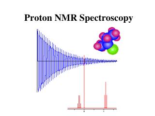
NMR Spectroscopy – Part 2 Judith Klein-SeetharamanDepartment of Structural Biology jks33@pitt.edu
NMR parameters Chemical Shift H2O methyl aromatic Trp-side-chain NH OH aliphatic Backbone NH Side-chain HN Ha Spectrum see handout Computational Biology Laboratory Course – Klein-Seetharaman – NMR Lecture
1d 1H NMR spectra Computational Biology Laboratory Course – Klein-Seetharaman – NMR Lecture
NMR of membrane proteins In detergent micelle: In lipid bilayer: http://www.elmhurst.edu/~chm/vchembook/558micelle.html Computational Biology Laboratory Course – Klein-Seetharaman – NMR Lecture
Problems! Detergent peaks Detergent signals cause dynamic range problems (Detergent signals cause spectral overlap) Detergent deuteration is often not feasible Problem: 1H,1H NOESY spectra do not show protein signals Computational Biology Laboratory Course – Klein-Seetharaman – NMR Lecture
Selective excitation B. Selective excitation of the same region as in A. Using excitation sculpting. A. Selective excitation of the NH region using 90 degree pulse followed by direct observation. Backbone NH Tryptophan side chain NH 20 15 10 5 10 5 0 -5 1H Chemical Shift [ppm] 1H Chemical Shift [ppm] Computational Biology Laboratory Course – Klein-Seetharaman – NMR Lecture
2d HSQC Computational Biology Laboratory Course – Klein-Seetharaman – NMR Lecture
1d projection of HSQC Computational Biology Laboratory Course – Klein-Seetharaman – NMR Lecture
HSQC spectra Computational Biology Laboratory Course – Klein-Seetharaman – NMR Lecture
Chemical shift perturbation Figure 2 in “Cap-free structure of eIF4E suggests a basis for conformational regulation by its ligands Laurent Volpon, Michael J Osborne, Ivan Topisirovic, Nadeem Siddiqui and Katherine LB Borden The EMBO Journal (2006) 25, 5138–5149 Computational Biology Laboratory Course – Klein-Seetharaman – NMR Lecture
Assignment is needed! Computational Biology Laboratory Course – Klein-Seetharaman – NMR Lecture
Several different assignment strategies exist Most easily automated: • HNCO • HNCOCACB • HNCOCA • HNCACB • HNCA • HNCACO Computational Biology Laboratory Course – Klein-Seetharaman – NMR Lecture
Experiments Figure 9. HNCO experiment. The magnetization is transferred (blue arrows) from the HN(i) proton via the N(i) atom to the directly attached CO(i-1) carbon atom and returns the same way to the HN(i) nucleus which is directly detected. The frequencies of all three nuclei (red) are detected. Image and description downloaded from http://www.cryst.bbk.ac.uk/PPS2/projects/schirra/html/3dnmr.htm Computational Biology Laboratory Course – Klein-Seetharaman – NMR Lecture
Experiments Figure 14. HNCA experiment. The HNCA experiment is the prototype of all triple resonance experiments. Starting at an amide proton (H) the magnetization is transferred to the directly attached nitrogen atom (N) which is measured as the first spectral dimension. Then the magnetization is transferred to the Calpha nucleus (CA) which is measured as second dimension. Afterwards, the magnetization is transferred back the same way to the amide proton which is measured as the third (direct) dimension. Image and description downloaded from http://www.cryst.bbk.ac.uk/PPS2/projects/schirra/html/3dnmr.htm Computational Biology Laboratory Course – Klein-Seetharaman – NMR Lecture
Experiments Figure 15. HNCACO experiment. In the HN(CA)CO experiment the magnetization is transferred from the HN(i) proton via the N(i) atom and the CA nucleus (Calpha(i)) to the CO(i) carbon atom and back the same way. The Calpha atom (yellow) acts only as relay nucleus, its frequency is not detected. It is only the frequencies of HN, N and CO (red) which are detected. Image and description downloaded from http://www.cryst.bbk.ac.uk/PPS2/projects/schirra/html/3dnmr.htm Computational Biology Laboratory Course – Klein-Seetharaman – NMR Lecture
Assignments Computational Biology Laboratory Course – Klein-Seetharaman – NMR Lecture
Structure Prediction by NMR Computational Biology Laboratory Course – Klein-Seetharaman – NMR Lecture
NMR parameters • chemical shifts • NOE • Dipolar coupling • Scalar coupling constants (gives dihedral angles) • Solvent exchange • HetNOE • longitudinal relaxation rates (R1) • transverse relaxation rates (R2) Computational Biology Laboratory Course – Klein-Seetharaman – NMR Lecture
NMR parameters The Nuclear Overhauser Effect http://www.oci.unizh.ch/group.pages/zerbe/NMR.pdf Computational Biology Laboratory Course – Klein-Seetharaman – NMR Lecture
HSQC TOCSY http://www.oci.unizh.ch/group.pages/zerbe/NMR.pdf Computational Biology Laboratory Course – Klein-Seetharaman – NMR Lecture
NMR Parameters Dipolar Couplings http://www.oci.unizh.ch/group.pages/zerbe/NMR.pdf Computational Biology Laboratory Course – Klein-Seetharaman – NMR Lecture
Scalar coupling constants Computational Biology Laboratory Course – Klein-Seetharaman – NMR Lecture
Structure Calculations • Distance geometry • Determines ensembles of structures consistent with an incomplete set of distance restraints • Metric matrix algorithm • Variable target function approach • Restrained molecular dynamics • Cartesian or torsion-angle coordinate systems • Molecular dynamics force fields are supplemented by pseudo energy terms based on the NMR-derived restraints Computational Biology Laboratory Course – Klein-Seetharaman – NMR Lecture
Structure Prediction From NMR Parameters Most widely used software suites • CNS • http://cns.csb.yale.edu/v1.1/ • XPLOR • http://xplor.csb.yale.edu/xplor/ Computational Biology Laboratory Course – Klein-Seetharaman – NMR Lecture
NMR parameters • chemical shifts • NOE • Dipolar coupling • Scalar coupling constants (gives dihedral angles) • Solvent exchange • HetNOE • longitudinal relaxation rates (R1) • transverse relaxation rates (R2) Computational Biology Laboratory Course – Klein-Seetharaman – NMR Lecture
Comparison of T1 and T2 relaxation http://www.oci.unizh.ch/group.pages/zerbe/NMR.pdf Computational Biology Laboratory Course – Klein-Seetharaman – NMR Lecture
Dynamics in folded/unfolded lysozyme Unfolded: Folded: Smaller rates – more flexible Computational Biology Laboratory Course – Klein-Seetharaman – NMR Lecture