Download
1 / 28
370 likes | 753 Views
2009. Gametogenesis. Gametes – reproductive cells. Ovum Spermatozoon Gametogenesis – differentiation of highly specialized sex cells capable of uniting at fertilization 1. Origin of the germ cell 2. Multiplication in the gonads by mitosis 3. Reduction of chromosomes – meiosis

E N D
2009 Gametogenesis
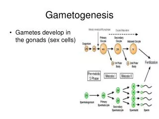
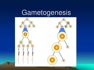
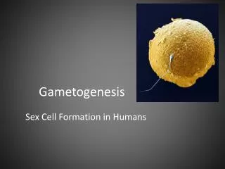
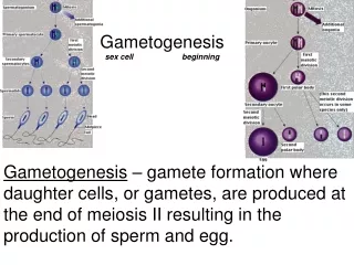
Gametes – reproductive cells • Ovum • Spermatozoon • Gametogenesis – differentiation of highly specialized sex cells capable of uniting at fertilization • 1. Origin of the germ cell • 2. Multiplication in the gonads by mitosis • 3. Reduction of chromosomes – meiosis • 4. Final stages of maturation and differentiation
Origin of primordial cells • Germ cell can be recognized very early – vegetal pole cytoplasm in the zygote • Epiblast – temporary residence in extraembryonic tissues – recognizable at 24 ED in the endoderm of yolk sac • Migration within mesenchyme of posterior wall of yolk sac (near the allantois), gut, and dorsal mesentery (4 -6 week) to the gonads • Extracellular matrix and chemotactic influence from gonad – resident germ cells induce formation of gonads • Number of cells increases during migration
Proliferation • Oogonia and spermatogonia • Proliferative phase of development – from thousands to about 7 million (in female) – mitosis • Oogonia – division during 2.-5. months • By the seventh month oogonia entere the prophase of first meiotic division and end proliferative phase • Spermatogonia enter meiosis after puberty, mitotic capability continues as long as the male is capable of reproduction
Meiosis • Reduction of normal number of chromosomes • From diploid to haploid • Two maturation divisions without new DNA synthesis • Reductional division • Equational meiotic division • Recombination of genetic information • Random distribution of maternal and paternal chromosomes • Exchanging of portions of homologous chromosomes by crossing over
First Meiotic Division • Prophase I • Leptotene • Zygotene • Pachytene • Diplotene • Diakinesis • Metaphase I • Anaphase I • Telophase I and Interphase • Second Meiotic Division
In Mammals - initiation of germ line development- maintain pluripotency within germ cells • Activation of differentiation – inductive signal from trophoblast • Proliferation and survival – trophic factors • Extracellular matrix – direct the migration • Final differentiation
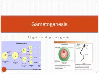
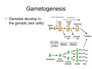
Spermatogenesis - 64 days • Mitotic multiplication – spermatogonia (Type A – stem cell population, Type B – leave mitotic cycle - preleptotene spermatocytes) • Meiosis - Primary spermatocytes • Secondary spermatocytes • Spermiogenesis – Spermatides – transformation into extremely specialized cells – spermatozoa (concentration of chromatin, decrease of size, formation of acrosome, flagellum)
Male germ cells • Sertoli cells – isolation of germ cells, support and nutrition • Degradation of residual bodies • Synthesis of signal molecules (Anti-Müllerian factor) • Synchronization of development- waves
Spermiogenesis • Nucleus – concentration of chromatin – head • Golgi complex- proacrosomal granules - acrosome • Centrioles – achorage of flagellum • Axoneme – microtubules (9+2) and dynein • Mitochondria – spiral investment around proximal part of flagellum – mitochondrial helix • Residual body
Spermatozoon • Head (nucleus and acrosome) • Neck (proximal centriole) • Middle piece (flagellum, centriole, mitochondrial helix) • Tail - flagellum
Sperm maturation • Newly formed spermatozoa are not capable of fertilization. Maturation in genital tract – activation – increase of motility • Capacitation – final step of sperm maturation- changes in acrosome, preparing the enzyme release (in female genital tract), changes in sperm membrane • Sperm attraction and hyperactivation • Acrosome reaction – fusion of the acrosome with plasma membrane, extension of the acrosomal process
Oogenesis • Oogonium gives arise to only one ovum – first and second polar body (DNA and only little cytoplasma) • First meiotic division is not completed untill puberty • Meoitic arrest occurs during prophase I (diplotene) – egg builds up its stores of yolk • Second arrest during metaphase II – mitosis is finished after fertilization
Lampbrush chromosomes • Active transcription during meiosis • Synthesis of RNA – genes loop out
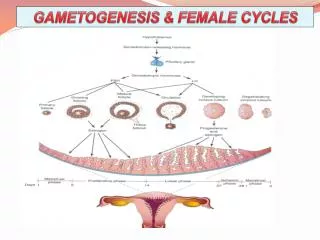
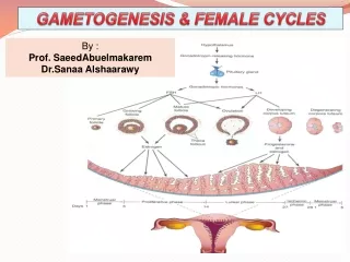
Oogenesis • At birth – 1 milion oocytes • Surrounded by a layer of follicular cells (granulosa cells) – follicle • Only 400 (one per menstrual cycle) reach maturity • Atresia (degeneration) • Folliculogenesis • Primordial • Primary • Secondary • Graafian follicle - Ovulation
Egg • Egg accumulates yolk as reservoir of food (energy) for embryo • Proteins (Amino acids, Energy) • Ribosomes and tRNA- proteosynthesis after fertilization • mRNA – early development - morphogenic factors
Coverings of eggs • Zona pellucida – Glycoproteins, GAG, Hyaluronic acid, Sialic adid. It is produced by oocyte • ZP-3 Sperm receptor and induction of acrosome reaction • Corona radiata – follicular cells
Fertilization • It is an interaction between sperm and oocyte • Spermatozoon binds to specific sperm receptor in the zona pellucida (ZP3). It induces release of enzymes from acrosome • Penetration the zona pellucida • Sperm and oocyte fuse • Cortical reaction – cortical granules release to perivitelline space (between oocyte and zona pellucida) – alteration of receptors for sperms – prevent polyspermy
Prevention of polyspermy • Fast block of polyspermy – change the electrical potential • Slow block of polyspermy - cortical granules -enzymes – proteases – clip off binding receptor • Fertilization envelope – space between zona pellucida and egg - GAG, peroxidase, and hyalin – zona reaction
Fertilization • Fusion with sperm induces oocyte to resume meiosis – second polar body and definitive oocyte • Fertilized oocyte = zygote • Female and male pronuclei • Membrane disapears • Replication • First mitotic division • 24 hours
Imprinting • Egg-derived genome is functionally different from sperm-derived • Imprinting is inactivation of gene depending on gender - prevent parthenogenesis • Maternal genes are important for embryo development (receptor for IGFII) • Paternal genes are important for placenta development (IGFII – Beckwith-Wiederman sy)
Cleavage • Mitotic division without cell growth • Daughter cells (Blastomeres) get smaller - embryo does not change in size • Mitotic division is equal and total • 4 cells – 40 hours • 3ED – 6-12 cells • 4ED – 16 -32 cells – morula (mulberry)
Segregation of blastomeres into embryoblast and trophoblast • Starting at 8 cell stage – changes in intercellular juctions – compaction – polarization of cells • Tight junction and gap junctions – outer cell mass • Cells in centre – inner cell mass (embryoblast) and outer cell mass – (trophoblast). • Fluid is collected – blastocyst cavity • Blastocyst – Embryonic pole • Abembryonic (vegetative) pole
Genetic regulation of germ cell formation, proliferattion, migration, and development • Regulatory gene cascade – sequential activation of genes that direct the initial induction and development, proliferation, survival, migration and differentiation of the germ cells • Maternal effect genes – germ plasm in zygote
Twins and embryonic stem cells • Monozygotic twins - before hatching – at 5.ED – dichorionic • Later monochorionic,diamniotic • Monochorionic monoamniotic • Conjoined twins (after ED9) • Inner cell mass – embryonic stem cells