Download
1 / 24
400 likes | 2.3k Views
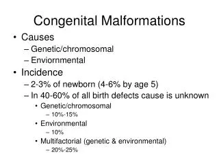
Hemangiomas and Vascular Malformations. Hemangiomas. Infantile Hemangiomas Most common vascular tumor of infancy 10% More common in Caucasians Females Premature infants Placental abnormalities Location >50% head and neck 25% trunk Rest on extremities Timing

E N D
Hemangiomas • Infantile Hemangiomas • Most common vascular tumor of infancy • 10% • More common in • Caucasians • Females • Premature infants • Placental abnormalities • Location • >50% head and neck • 25% trunk • Rest on extremities • Timing • Several days to weeks after delivery
Hemangiomas • Infantile Hemangiomas • Description • Reddening or bluish discoloration of skin • Bright red nodule or plaque with elevation • Types • Superficial (epidermal) • “Strawberry” or bright red • Well demarcated • Elevated • Soft compressible • Few mm to 5cm • Deep (dermis or subq fat) • Bluish hue • Indistinct borders • Doughy consistency • Enlarge when dependent • Mixed • Most hemangiomas
Hemangiomas • Infantile Hemangiomas • Course • Grow and peak by 6-9 months • Stabilization • Involution • 10% per year • Graying out of surface • 40% with residual skin changes • Telangiectasias • Fibro-fatty tissue
Hemangiomas • Hemangiomatosis • Multiple hemangiomas • Benign • Limited to the skin • Not benign • Numerous small (<2cm), widely dispersed cutaneous lesions • Internal or visceral lesions • Liver • May have AV shunts and precipitate high-output CHF • 6-12 weeks of age • GI tract • Bleeding • CNS • Mass effect • Lungs
Hemangiomas • When to worry? • Lower face • Lower lip, chin, preauricular, neck • “beard” distribution • Airway involvement • Midline lumbosacral • Spinal dysraphism
Hemangiomas • PHACES syndrome • Posterior fossa malformations • Hemangiomas • Plaque-like segmental hemangioma of the face • May initially be confused with port-wine stain • Often ulcerate and proliferate rapidly • Arterial anomalies • Carotid • Cardiac defects • Eye anomalies • Sternalclefting
Hemangiomas • Complications • Periorbital and lid lesions • Occlusion of the visual axis • Corneal compression • Must be treated aggressively • Amblyopia, strabismus, astigmatism • Lips, nose or ears • High potential for disfigurement • High friction areas • Ulceration • Secondary infection • Scarring
Hemangiomas • Treatment • Conservative management for most • Lesions involving the airway or the eye • Steroids • Interferon • Surgical intervention
PyogenicGranuloma • Common benign vascular tumors • Overgrowth of granulation tissue • Following minor trauma • Foreign body • Timing • Well after the newborn period • Location • Usually face or extremity • Description • Solitary bright red, soft nodules • Pedunculated • 5-6mm • Friable surface • Treatment • Excision • Electrodessication of the “feeder” vessels • May recur
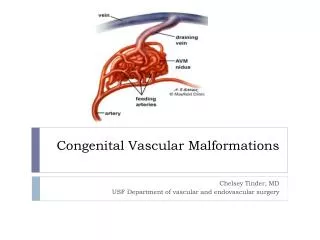
Vascular Malformations • Nevus Simplex • AKA Salmon Patch or “Stork Bite” • Capillary malformation • Seen in majority of infants at birth • Location • Nape of neck • Glabella • Forehead • Upper eyelids • Lower back • Course • Fade with time • More apparent when crying or straining
Vascular Malformations • Nevus Flammeus • AKA Port-Wine stain • Congenital capillary/venous malformation • Description • Purple-red • Location • Unilaterally on face • Course • Do not enlarge or involute
Vascular Malformations • Sturge-Weber Syndrome • Port wine stain • Distribution of the trigeminal nerve • Vascular malformations of the ipsilateralleptomeninges and cerebral cortex • Glaucoma • Other • Seizures, MR, hemiplegia • Klippel-Trenaunay Syndrome • Port wine stain • Over an extremity • Hemihypertrophy • Soft tissue and bony overgrowth
Question 9 A new adolescent patient is seen in your office for a sports physical. He points out a hairless, well-circumscribed, yellowish waxy plaque located on his scalp. He says its been there since birth but has recently become more raised. He wants to know what it is? A. Epidermal nevi B. Congenital nevomelanocytic nevi C. Halo nevus D. Nevus sebaceous E. Ash-leaf spot
Nevi • Congenital Nevomelanocytic Nevi • Description • Pigmented plaques often associated with dense hair growth • Course • Birth • Tan or light pink with soft vellus hairs • Infancy and childhood • Darkening with small dark macules or nodules within the plaque and prominent hair
Nevi • Congenital Nevomelanocytic Nevi • Size • Small <1.5cm • Medium 1.5-20cm • Large or Giant >20cm • Prognosis • All have potential for malignant transformation • New, darker and/or bleeding nodules • Sudden growth • 1-4% small to medium • 10-30% Giant
Nevi • Congenital Nevomelanocytic Nevi • Management • Small to medium • Yearly follow-up with derm • Excision if atypical or difficult to monitor • Giant • Early, full thickness excision followed by grafting OR • Close observation every 6 months
Nevi • Acquired Nevomelanocytic Nevi • Timing • Early childhood • Description • Small, flat, pigmented macules • 1-2mm • Location • Sun-exposed areas • Course • Junctional nevi • Limited to epidermal-dermal junction • Compound nevi • Papular or pedunculated • Proliferation into the dermis • Change slowly over months and only warrant observation
Nevi • Acquired Nevomelanocytic Nevi • Halo nevus • Hypopigmented or depigmented ring associated with mild local pruritus around a benign nevus • Caused by cytotoxic T-lymphocyte reaction • Course • Eventual resolution and nevus disappears
Melanomas • Childhood • De novo • Within giant congenital nevus or other nevus • Transplacental transfer • Red Flags • Change in size, shape or outline • Scalloped, irregular borders • Change in surface characteristics • Small, dark, elevated papule or nodule within a flat plaque • Flaking, scaling, ulceration or bleeding • Change in color • Different shade or to a mixture of red, white or blue • Development of burning, itching or tenderness
Melanoma Differential • Blue nevus • Small, firm, blue papule • Deep nevus cells • Traumatic hemorrhage • Under the nails or in mucous membranes • Vascular lesions • Pyogenicgranuloma or angiokeratoma • Spitz nevus • Red and rapidly growing nevus • Composed of spindle and epithelial cells • Confused histologically with melanoma
Hamartomatous Nevi • Epidermal Nevi • Epidermal structures only • Timing • Birth or childhood • Description • Slightly hyperpigmentedpapillomatous or verrucous growth • Increases in verrucous changes are common at puberty • Small and localized, linear, dermatomal or generalized • Other associations if extensive • Seizures, MR, ocular and skeletal defects, hypophosphatemic vitamin D-resistant rickets
Hamartomatous Nevi • Nevus sebaceous of Jadassohn • Epidermal proliferation, abortive hair follicles, sebaceous glands and apocrine structures • Timing • Present at birth • More verrucous, raised and nodular at puberty • Description • Hairless, well-circumscribed, skin-colored or yellowish waxy plaque • Linear or round • Location • Scalp, face or neck • Course • Routine excision not recommended in childhood • Malignancy risk is low • May excise in adolescence or adulthood