Download
1 / 65
670 likes | 884 Views
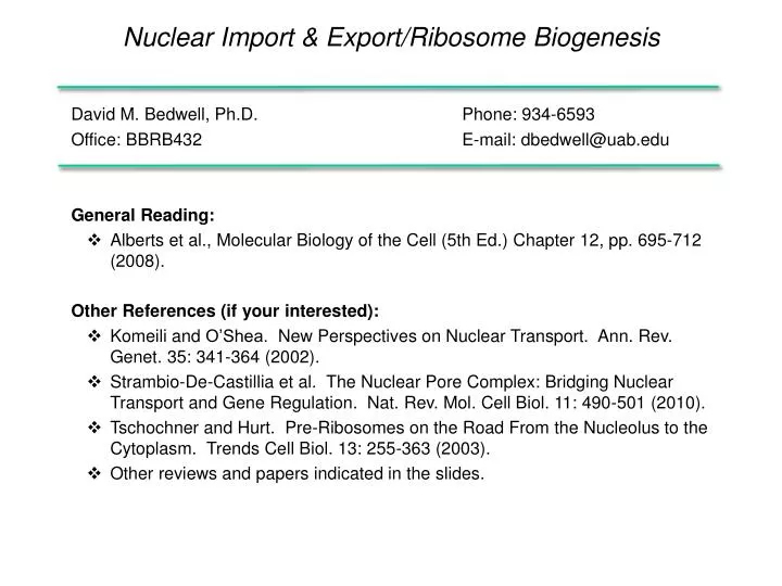
Nuclear Import & Export/Ribosome Biogenesis. David M. Bedwell, Ph.D. Phone: 934-6593 Office: BBRB432 E-mail: dbedwell@uab.edu General Reading : Alberts et al., Molecular Biology of the Cell (5th Ed. ) Chapter 12, pp. 695- 712 (2008). Other References (if your interested):

E N D
Nuclear Import & Export/Ribosome Biogenesis • David M. Bedwell, Ph.D. Phone: 934-6593 • Office: BBRB432 E-mail: dbedwell@uab.edu • General Reading: • Albertset al., Molecular Biology of the Cell (5th Ed.) Chapter 12, pp. 695-712 (2008). • Other References (if your interested): • Komeiliand O’Shea. New Perspectives on Nuclear Transport. Ann. Rev. Genet. 35: 341-364 (2002). • Strambio-De-Castillia et al. The Nuclear Pore Complex: Bridging Nuclear Transport and Gene Regulation. Nat. Rev. Mol. Cell Biol. 11: 490-501 (2010). • Tschochner and Hurt. Pre-Ribosomes on the Road From the Nucleolus to the Cytoplasm. Trends Cell Biol. 13: 255-363 (2003). • Other reviews and papers indicated in the slides.
Lecture Overview • Overview of cellular compartmentalization. • Features of nuclear pores. • Mechanism of nuclear Import. • Mechanism of nuclear export. • Ribosome assembly and export.
Protein Trafficking Mechanisms • There are two basic pathways of biosynthetic protein traffic: • Default localization • Signal-mediated localization • gated transport • transmembrane transport • vesicular transport
Intracellular Protein Transport Mechanisms Cytosol Nucleus Peroxisome Mitochondria Plastids gated transmembrane vesicular Endoplasmic Reticulum Golgi Apparatus Lysosome Late Endosome Secretory Vesicles Early Endosome Cell Surface
Three-Dimensional Model of the Nucleus Nuclear Architecture
Scanning EM of the Nucleus Blue pseudo-coloring highlights the nuclear pore complexes, while green pseudo-coloring highlights the nuclear envelope together with the attached ribosomes. Kiseleva, Nature Cell Biol. 6: 497 (2004)
Types of Traffic That Pass Through Nuclear Pores Imported RNA Polymerases snRNPs DNA Polymerases Ribosomal Proteins Histones Transcription factors Exported 40S ribosomal subunits 60S ribosomal subunits tRNAs mRNAs snRNAs
Volume of Traffic Through Nuclear Pores • A single HeLa cell contains 10 million ribosomes, ~4000 nuclear pores, and divides every 24 hrs. This means a total of: • 400,000 ribosomal proteins must be imported each minute (~100 r-proteins/pore). • 12,000 ribosomal subunits must be exported each minute (~3 ribosomal subunits/pore). • If synthesizing DNA, need ~1 million new histone molecules every 3 minutes, so need to transport 100 histones/pore each minute. • Several hundred other proteins, RNAs, and RNPs move in and out of a single nuclear pore each minute.
Nuclear Pore Complex • 8 fold rotational symmetry. • Size exclusion ranges from 9 nm (“closed”) to 26 nm (“open”).
Nuclear Pores Embedded in the Nuclear Membranes The nuclear pore contains spoke and ring assemblies that are integrated into the two membranes of the nuclear envelope.
Nuclear Pore Complex (NPC) Composition • A single nuclear pore contains ~ 1000 proteins (total) and 60-100 different proteins. These nuclear pore complex (NPC) proteins are called nucleoporins. • Many nucleoporins are glycoproteins that carry O-linked N-Acetylglucosamine (Glc-NAc) residues. • Nuclear pore fibrils and other nucleoporins within the NPC channel contain phenylalanine and glycine (FG) repeats that facilitate binding of the nuclear import receptors to the nuclear pore complex during its translocation through the nuclear pore. These interactions allow transported molecules to pass bi-directionally through the 15nm long pore. • A relative size perspective: The NPC has a MW of 125 million Daltons. By comparison, a mammalian ribosome has a molecular weight of ~ 4 million Daltons.
Nuclear Pore Complex Structure • Each nuclear pore complex (NPC) is a cylindrical structure comprised of eight spokes surrounding a central tube that connects the nucleoplasm and cytoplasm. • The outer and inner nuclear membranes (ONM and INM, respectively) of the nuclear envelope join to form grommets in which the NPC sits. • The NPC is anchored to the nuclear envelope by a transmembrane ring structure that connects to the core scaffold and comprises inner ring and outer ring elements. • Linker nucleoporins (Nups) help anchor the Phe-Gly (FG) Nups such that they line and fill the central tube. Strambio-De-Castillia et al., Nat. Rev. Mol. Cell Biol. 11: 490-501 (2010)
The Nuclear Pore Complex Functions as a “Virtual Gate” • The outer and inner nuclear membranes (ONM and INM, respectively) of the nuclear envelope join to form a ring-shaped pore where the nuclear pore complex (NPC) resides. • At the NPC, the nucleus and cytoplasm are connected by a channel, which is filled with flexible, filamentous Phe-Glynucleoporins (FG Nups). • Spurious macromolecules are physically excluded from entering the densely packed FG Nup meshwork. • Nuclear transport factor (NTF)-bound cargo can enter the channel from either its cytoplasmic or nucleoplasmic side and hop between binding sites on the FG Nups until they return to the original compartment or reach the opposite side of the NPC. Strambio-De-Castillia et al., Nat. Rev. Mol. Cell Biol. 11: 490-501 (2010)
Two Models For Natively Disordered FG-Repeat Domains in the Transport Channel of the Nuclear Pore Left: FG-repeat network may form a hydrogel, crosslinked by hydrophobic interactions between the phenylalanines. Right: FG repeats could form a network of unlinked polymers whose thermally activated undulations create a zone of "entropic exclusion”. Elbaum, Science 314: 766-767 (2006)
FG Repeats Can Form an Elastic Hydrogel in Aqueous Solution Left: An aqueous solution with 26 mg/ml wild-type FG-repeat domain from Nsp1p (400 µM) was filled into a silicon tubing, where it completed gelling. The formed gel was pushed out of the tubing by gentle pressure, placed onto a patterned support (1 square = 1.4 mm2), and photographed. Note that the pattern shows clearly through this transparent gel. Inset illustrates how interactions between the hydrophobic clusters (shown in red) cross-link the repeat domains into a hydrogel. The FG repeats can form a free-standing gel, and they measure elasticity comparable to 0.4% agarose. Right: The FS mutated repeat domain remained liquid after identical treatment. Frey et al., Science 314: 815-817 (2006)
Hydrogel Model of Nuclear Pore Function • Selective phase model for the passage of a nuclear transport receptor (NTR) through the permeability barrier of nuclear pore complexes. • Inter-repeat contacts between the hydrophobic clusters ( ) of FG-repeat-domains create a sieve-like barrier which restricts the passage of inert objects larger than the mesh-size. • NTRscan overcome this size-limit, because they possess binding sites ( ) for the hydrophobic clusters. They compete with inter-repeat contacts, thereby open adjacent meshes and dissolve within the barrier. Since the involved interactions are of low affinity, the NTR can leave the barrier on the other side. Frey et al., Science 314: 815-817 (2006); Burke, Science 314: 766-767 (2006)
Selective (Signal-Mediated) Nuclear Entry Nuclear pores don't close completely - time required for proteins that lack a nuclear targeting signal to diffuse through the nuclear pore in living cells has been measured: <5kD -seconds 17kd -2 minutes 44kD -30 min >60kD -does not enter nucleus Remarkably, even 20 nm gold particles coated with molecules having nuclear import or export signals can pass readily through the nuclear pore.
Outcomes of Nuclear Pore Function * * * * * * * * * * * * * * * N N N * * * * * * * * * * * * * * * * * * * * * * * * * * * * * * * * * Nuclear Exclusion Nuclear Localization Diffusion-Limited Equilibration
Characteristics of Nuclear Transport • Active transport through the nuclear pore complex (NPC) has the following features: • Energy dependent • Temperature dependent • Signal dependent • Saturable • These are features of a carrier-mediated process.
Nuclear Localization Signals • Two types of Nuclear Localization Signal (NLS): • Short basic sequences of 4-8 residues • [PPKKKRKV is the NLS of SV40 large T antigen] • Bipartite signals with two stretches of basic amino acids separated by ten less-conserved amino acids. • [KRPAATKKAGQAKKKK is the NLS of nucleoplasmin] • Both types of NLS are rich in the basic amino acids arginine and lysine and usually contain proline.
NLS Location of Nuclear Localization Signals Proteins don’t unfold during nuclear import. An NLS can be located anywhere in a protein, as long as they lie on the surface of the folded protein molecule where they can be recognized by an NLS receptor.
Methods Used to Identify NLSs • Microinjection studies- can be used to study nuclear targeting signals either in their natural context, when fused to passenger proteins, or when stuck to gold particles. • Deletion and gene fusion studies- Deletions can be used to identify regions necessary for nuclear import, while the fusion of these sequences to a passenger protein tests whether these sequences are sufficient for nuclear import. • Mutational analysis- Determine specific amino acid sequence necessary for nuclear localization.
Use of Electron Microscopy to Identify NLSs Electron micrograph showing nuclear entry of colloidal gold particles coated with nucleoplasmin following microinjection.
Use of Immunofluorescence to Identify NLSs The 8 amino acid SV40 NLS can target a cytosolic protein to the nucleus when introduced either genetically or by crosslinking. Pyruvate Kinase plus SV40 NLS Pyruvate Kinase
Mutational analysis of the SV40 Large T antigen (90 kDa protein required for viral DNA replication)
Mechanism of Nuclear Import • Importin-, a component of the nuclear localization signal (NLS) receptor complex binds to the NLS of a protein to be imported. • Importin-, the other subunit of the NLS receptor complex, mediates docking with the outer surface of the nuclear pore in a rapid, energy-independent fashion. • Translocation of the trimeric complex occurs along FG-repeat proteins within the nuclear pore in an energy-dependent manner. Importin- interacts with the FG-containing components of the pore complex. • Once the complex enters the nucleoplasm, Ran-GTP binds, releasing the cargo molecule from the complex. • Following the dissociation of the imported protein from the complex, the receptor components (with bound Ran-GTP) are then re-exported to the cytoplasm for another cycle.
Mechanism of Nuclear Import (cont) • Three important accessory proteins assist Ran function: • A cytosolic Ran Binding Protein (BP) dissociates Ran-GTP from the receptor. • The cytosolic Ran GTPase-Activating Protein (Ran-GAP) triggers GTP hydrolysis, converting Ran-GTP to Ran-GDP. • The Ran Guanine nucleotide Exchange Factor (Ran-GEF), which promotes exchange of GDP to GTP, is nuclear. • The nuclear location of the Ran-GEF maintains nuclear Ran in the GTP-bound form, providing directionality to nuclear transport. • Once cytosolic Ran-GTP is hydrolyzed to Ran-GDP by Ran-GAP, the Ran-GDP is then re-imported into the nucleus for another cycle.
Regulated Nuclear Import • In some cases, pre-synthesized transcription factors and cell cycle regulators are maintained in the cytoplasm and only translocate into the nucleus at specific times or in response to specific signals. • Mechanisms used to achieve regulated entry include: • Aconformational change upon ligand binding. • Covalent modification (e.g., phosphorylation of NLS). • Attachment to a cytoplasmic structure to block import. • Binding of regulatory subunits that mask the NLS.
Nuclear Transport Ligand-Induced Activation of an NLS NLS NLS Ligand-Induced Conformational Change Nucleus Cytosol
PO4 NLS NLS NLS Regulation by Covalent Modification Dephos-phorylation Nuclear Transport Nucleus Cytosol
Regulation by Covalent Modification (cont) • NFAT (Nuclear Factor of Activated T cells) is a transcription factor that contains a nuclear localization sequence, but it is buried in the protein interior. • Whether NLS or NES is masked depends on the phosphorylation state of specific serine residues in the regulatory domain. Phosphorylation of these serine residues exposes an NES, whereas dephosphorylation exposes an NLS. • In resting cells, the NLS of the cytoplasmic NFAT is masked due to phosphorylation on these serine residues. • In stimulated cells, an increase of intracellular calcium ions activates calcineurin, which then dephosphorylates the masking residues. Consequently, the NLS is exposed and NFAT can be carried into the nucleus by the importin/ complex. • Inside the nucleus, NFAT may be re-phosphorylated by a protein kinase, exposing its NES so it can be exported by the exportin Crm1.
Induced Activation of Nuclear Entry by the Level of Cytosolic Calcium Nuclear entry of the transcription factor NFAT is induced when the level of cytosolic calcium increases.
NLS NLS NLS Release Nuclear Transport Regulation by Cytosolic Retention Nucleus Cytosol Cytoskeletal Elements
NLS Masking by a Regulatory Subunit Glucocorticoid Receptor
Why aren’t nuclear localization signals removed following import?
The Lamina Controls Nuclear Integrity • At the onset of mitosis, phosphorylation of nuclear lamins leads to the dissassembly of the lamina and the subsequent breakdown of the nuclear membrane. • Prior to nuclear re-assembly, dephophorylation of the lamins occurs. LaminB, which remains associated with a specific receptor on nuclear membrane vesicles, is then rejoined by lamins A and C. This is followed by the reassembly of the lamina and the membrane in a GTP-dependent process. • Mutations in the gene encoding lamin A have been shown to be associated with at least six different diseases that are collectively called the laminopathies.
Repeated Nuclear Entry • Nuclear proteins are capable of repeated entry into the nucleus because nuclear localization signals are not removed when the protein enters the nucleus. • This is important, because when the cell undergoes mitosis, the nuclear membranes break down and nuclear proteins freely mix with cytosolic proteins. • Once mitosis is completed and the nuclear membranes re-form the nuclear proteins are imported again. This process can occur repeatedly.
Features of Nuclear Export • Nuclear export occurs by a mechanism analogous to nuclear import: • Protein to be exported contains a leucine-rich Nuclear Export Signal (NES). • A substrate to be exported is bound by an export receptor (such as Crm1) and Ran-GTP mediates its export from the nucleus. • Once in the cytosol, Ran-BP dissociates the exported substrate and its receptor. • Ran-GAP converts Ran-GTP to Ran-GDP. • Ran-GDP and the export receptor are then re-imported into the nucleus for another cycle of export.
CRM1-Mediated Nuclear Protein Export • (a) The CRM1 transport cycle. • In the nucleus, Ran-GTP stimulates binding of CRM1 to NES substrates. • After passage through the NPC, the CRM1/Ran-GTP/NES substrate complex is disassembled at the cytoplasmic filaments by the concerted action of Ran-BP1 and Ran-GAP. • The NES substrate is released to the cytoplasm and empty CRM1 is recycled back to the nucleus. • (b) Model of CRM1 export complex disassembly. • CRM1 is released into the cytoplasm and, for recycling into the nucleus, binds to a series of different, cargo-independent CRM1-binding sites. Kutay and Güttinger, Trends Cell Biol. 15: 121-124 (2005)
† † † PKA PKA Nucleus PKA † † PKA PKA † PKA † † † PKA PKA PKA Example of Nuclear Protein Export If you inject Protein Kinase A (PKA) and PKA Inhibitor (PKI)(†) into a cell nucleus, the PKI binds to PKA and transports PKA out of the nucleus by an active mechanism. Many proteins and RNAs undergo export from the nucleus. Nuclear Export Signals (NES) mediate the export of protein and RNA species. PKI transport of PKA out of the nucleus is both temperature and energy dependent, indicating an active process. PKI contains a Nuclear Export Signal (NES) [LALKLAGLDI]
Nuclear Export of Various RNA Species • mRNAs, snRNAs and ribosomes are transported in or out of the nucleus as ribonucleoprotein complexes (RNPs). • Like protein transport, RNA transport is signal-dependent, carrier mediated, and occurs through the nuclear pore complex. • In general, nuclear export is mediated by adaptor proteins and export receptors (exportins). • Adaptor proteins bind the export signal and present it to the exportin, which facilitates transport of the complex through the nuclear pore complex. • However, different classes of RNAs utilize different adaptors and receptors. Not all require Ran-GTP.