Download
1 / 28
280 likes | 503 Views
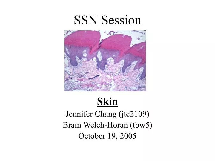
SSN Session. Skin Jennifer Chang (jtc2109) Bram Welch-Horan (tbw5) October 19, 2005. Functions. Protection - barrier to outside environment abrasion, moisture, UV light, microorganisms Homeostasis - maintenance of internal environment temperature, water and salts Sensation

E N D
SSN Session Skin Jennifer Chang (jtc2109) Bram Welch-Horan (tbw5) October 19, 2005
Functions • Protection - barrier to outside environment • abrasion, moisture, UV light, microorganisms • Homeostasis - maintenance of internal environment temperature, water and salts • Sensation • touch, temperature, pain, pressure, vibration • Immune surveillance • Langerhans’ cells – type of macrophage • Endocrine function • synthesizes vitamin D
Glabrous Hairy Types of Skin Thick – covers palms of hands and soles of feet, glabrous vs. Thin – covers rest of body, mostly hairy
3 layers • Epidermis stratified squamous keratinizing epithelium • Dermis papillary layer of loose connective tissue underlain by dense irregularly arranged CT • Hypodermis aka subcutaneous tissue, loose CT, contains adipose tissue
Epidermis – stratified squamous keratinized epithelium 5 Layers 1. Stratum corneum 2. Stratum lucidum 3. Stratum granulosum 4. Stratum spinosum 5. Stratum basalis
Stratum corneum Superficial keratinized layer Cells have no nuclei or organelles Sealed extracellular space Most superficial cells are sloughed off Stratum granulosum Basophilic granules of keratohyalin Promotes aggregation of keratin filaments into tonofibrils Lamellar bodies – water barrier Stratum corneum
Stratum Spinosum desmosomes • several cell layers thick • attached by intercellular bridges (desmosomes) • cells artificially pulled apart, the attachment sites give spiny appearance • Langerhans’ cells found here
mitotic cell cells with melanin Stratum Basalis • Mitotic cell layer, attaches to basement membrane via hemidesmosomes • Cells containing melanin may be either melanocytes or keratinocytes • Melanin • - pigment - protection from UV rays • - synthesized in melanocytes using tyrosinase - taken up by keratinocytes
Question 1 In the stratum basalis, ______ are found. They attach keratinocytes to ______. • Hemidesmosomes, basal lamina • Desmosomes, basal lamina • Hemidesmosomes, other keratinocytes • Desmosomes, Langerhans cells Lab 8, slide 2
Question 1 In the stratum basalis, ______ are found. They attach keratinocytes to ______. • Hemidesmosomes, basal lamina • Desmosomes, basal lamina • Hemidesmosomes, other keratinocytes • Desmosomes, Langerhans cells Lab 8, slide 2
Question 2 • Which of the following regarding these cells is true? • They are mitotic • They do not have organelles • They contain keratohyalin granules • They synthesize melanin
Question 2 • Which of the following regarding these cells is true? • They are mitotic • They do not have organelles • They contain keratohyalin granules • They synthesize melanin
Question 3 • This brown substance is synthesized in which cell layer? • Stratum corneum • Stratum granulosum • Stratum spinosum • Stratum basalis
Question 3 • This brown substance is synthesized in which cell layer? • Stratum corneum • Stratum granulosum • Stratum spinosum • Stratum basalis
Specialized Structures in Skin • Nerve supply • Hair follicles • Sweat glands • Sebaceous glands • (Nails) • (Mammary glands) (http://www.columbia.edu/~johan/images/arm.jpg)
The Dermis • Reticular layer • dense CT (less cellular) • eccrine sweat glands • Pacinian corpuscles • anatomy: Langer’s lines • Papillary layer • loose CT • bv’s, nerves, lymphatics • papillae into epidermis • Meissner’s corpuscles (Ross, 4/e, p. 425)
Cutaneous Nerve Endings • (Free nerve endings) • pain & temperature • (Merkel’s cells) • high-res. tactile sensation (in stratum basale) • Meissner’s Corpuscles • touch • Pacinian Corpuscles • vibration & pressure (Skin lab, slide 16; Cajal stain)
Meissner’s Corpuscles • in dermal papillae (papillary layer of dermis) • mechanoreceptors • 2-point discrimination • encapsulated • CT capsule • contrast w/ free nerve endings (Ross, 4/e, p. 412)
Pacinian Corpuscles (Ross, 4/e, p. 431)
Pacinian Corpuscles • in deeper dermis & hypodermis • reticular layer • vibration & pressure • encapsulated • characteristic appearance • “onion” / “bull’s eye” • diagnostic for dermis • or hypodermis • hypodermis • loose CT, bv’s, fat, etc. • a.k.a., subcutaneous tissue, superficial fascia (Skin lab, slide 18)
Hair Follicles • epithelially derived • epidermal invagination • matrix of follicle equiv. to stratum basale • most of body surface • body temp. regulation • sebaceous glands • secrete into follicle • arrector pili • smooth muscle • “goosebumps” • assists gland secretion (Ross, 4/e, p. 414)
Sebaceous Glands • associated w/ hair follicle • secrete sebum between shaft & follicle • holocrine secretion • oil-filled cells apoptose • secretory product & cell debris discharged from gland • sebum may be protective • but is involved in acne (Skin lab, slide 21)
Eccrine Sweat Glands • coiled tubular glands in dermis (reticular layer) • not associated with hair follicles; widely distributed • secretory portion (w/ basal lamina) • myoepithelial cells • contract expel sweat • ducts – cuboidal cells • stratified (2 layers) • corkscrew path • reabsorb H2O, salt • sweat is hypotonic • contains H2O, salt, IgA • temp. regulation (Skin lab, slide 13)
Sweat Ducts (Skin lab, slide 12)
Question 4 • What is the function of this nerve ending, and which skin layer is it in? a) pain; hypodermis b) temp.; reticular layer c) two-pt. discrimination; papillary layer d) itch; epidermis (Skin lab, slide 15)
Question 4 • What is the function of this nerve ending, and which skin layer is it in? a) pain; hypodermis b) temp.; reticular layer c) two-pt. discrimination; papillary layer d) itch; epidermis (Skin lab, slide 15)
Question 5 • The product of the gland in this image would most likely be: a) attractive to a potential mate b) hypotonic c) mainly water d) oily and associated with cell debris (Skin lab, slide 23)
Question 5 • The product of the gland in this image would most likely be: a) attractive to a potential mate b) hypotonic c) mainly water d) oily and associated with cell debris (Skin lab, slide 23)