Download
1 / 39
480 likes | 814 Views
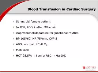
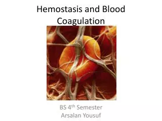
Coagulation, Fluid, and Blood Management for Cardiac Surgery. Maureane Hoffman, MD, PhD Professor of Pathology, Duke University and Director, Transfusion Service and Hematology Laboratory Durham Veterans Affairs Medical Center Durham, NC. Blood Coagulation and Lack Thereof …….

E N D
Coagulation, Fluid, and Blood Management for Cardiac Surgery Maureane Hoffman, MD, PhD Professor of Pathology, Duke University and Director, Transfusion Service and Hematology Laboratory Durham Veterans Affairs Medical Center Durham, NC
Blood Coagulationand Lack Thereof…… A topic which may be of concern during cardiac surgery
Objectives Appreciate differences between the “coagulation cascade” and how hemostasis works in vivo Understand that the PT and aPTT provide information about coagulation factor levels, but don’t necessarily reflect bleeding risk Consider approaches to restoring hemostasis in bleeding patients
There are reasons why many people don’t want to hear a talk about blood coagulation
Our group has been trying to develop better models to help us understand coagulation
In the 1960’s the coagulation factors were organized into a “cascade” or “waterfall” model. This evolved into the current cascade model … Macfarlane RG. An enzyme cascade in the blood clotting mechanism, and its function as a biological amplifier. Nature. 1964;202:498-499. Davie EW, Ratnoff OD. Waterfall sequence for intrinsic blood clotting. Science. 1964;145:1310-1312
The Coagulation Cascade aPTT PT Intrinsic Pathway Extrinsic Pathway FXII/HMK/PK Factor XI Factor XIa Factor VIIa Factor IXa Factor IX Tissue Factor Lipid Factor VIIIa Lipid Factor Xa Factor X Factor X Factor Va Lipid Thrombin Prothrombin Fibrinogen Fibrin
The “cascade” was intended as a model of how the coagulation proteins interact biochemically, not how hemostasis works in the body It IS a good model of what happens in the PT and aPTT assays
Intrinsic Pathway Prolonged aPTT only Factor XII/HMK/PK Prolonged aPTT Variable bleeding Factor XI FactorXIa Prolonged aPTT Severe bleeding Factor IXa Factor IX Factor VIIIa Factor X Factor Xa Factor Va Prothrombin Thrombin Fibrinogen Fibrin
Can putting the cells back in the model explain some clinical phenomena that the “protein-centered” cascade model cannot?
Cell-based conceptual model - Hemostasis occurs on two surfaces: TF-bearing cells and platelets 1.Initiation TF-Cell IIa 3.Propagation 2.Amplification Activated Platelet IIa Platelet
Initiation TF TF TF II X Xa VIIa VIIa Va IIa TF-Bearing Cell VIIa IX IXa Hoffman & Monroe: A Cell-Based Model of Hemostasis. Thromb Haemostas, 85:958-65, 2001
Amplification X Xa VIIa VIIa Va TF-Bearing Cell VIIa TF TF TF VIIIa XIa Va Activated Platelet II VIIIa + vWF VIII/vWF XI IIa Platelet V XIa V Va Priming Amount of Thrombin Hoffman & Monroe: A Cell-Based Model of Hemostasis. Thromb Haemostas, 85:958-65, 2001
Propagation TFPI Xa Xa VIIa VIIa Va TF-Bearing Cell VIIa TF TF TF VIIIa + vWF VIII/vWF XI IIa Platelet V XIa V Va II IX X Large amount of thrombin IXa Xa IX IIa VIIIa XIa XIa Va ActivatedPlatelet Hoffman & Monroe: A Cell-Based Model of Hemostasis. Thromb Haemostas, 85:958-65, 2001
A Cell-Based Model of Hemostasis II X VIII/vWF TF VIIa Xa IIa Va VIIIa TF-Bearing Cell TF XI XIa V Va VIIa IX Platelet II IXa X IIa Xa VIIIa IXa Va XIa Activated Platelet IX Hoffman M, et al. Blood Coagul Fibrinolysis. 1998;9(suppl 1):S61-S65.
There Really Are “Intrinsic” and “Extrinsic” Pathways • They are not redundant - they operate on different cellular surfaces to fill different roles • The “extrinsic” or TF pathway works on the initiating cells • The “intrinsic” pathway works on platelets to produce the thrombin “burst”
The extrinsic pathway acts in vivo to initiate coagulation PT Assay in vivo Adapted from: Monroe DM and Hoffman, M: What does it take to make the perfect clot? Arterio Thromb Vasc Biol 26:41-48, 2006
The intrinsic pathway acts on platelet surfaces to generate large amounts of thrombin in vivo aPTT Adapted from: Monroe DM and Hoffman, M: What does it take to make the perfect clot? Arterio Thromb Vasc Biol 26:41-48, 2006
Why do previously normal patients bleed? • Anatomic defects - “surgical” bleeding • Microvascular bleeding • Dilution or depletion of coagulation factors and platelets • Hyperfibrinolysis • Hypothermia • Acidosis • ?
Good News! The tests we have are fine for evaluating the cause of bleeding • The PT and aPTT are useful if we have a bleeding patient and we want to figure out if a factor deficiency is responsible
The tests we have are also fine for directing component therapy • Prolonged PT or aPTT = plasma • Low fibrinogen = cryoprecipitate • Low platelet count or defect on platelet function testing = platelet concentrates
The whole idea is to get a stable platelet/fibrin clot IIa Fibrinogen
Blood component therapy does not always stop the bleeding • Component therapy is generally intended to replace deficient factors/platelets • Replacement doesn’t always work • FFP is always somewhat diluted • Platelets have a “storage defect” • Even if we apparently restore “normal” levels, bleeding may not stop • Can exacerbate acidosis, hypocalcemia, hyperkalemia and hypothermia
What else can we do for microvascular bleeding? • Blood component replacement • Anti-fibrinolytics • Effective in some settings • Coagulation factor concentrates - can achieve supra-normal levels of factors • Recombinant FVIIa • Fibrinogen concentrate • Prothrombin complex concentrates
Fibrin clot structure depends on the amount/rate of thrombin generation and the amount of fibrinogen incorporatedHigher levels of each give more structurally stable clots
We can enhance thrombin generation by: • Replacing deficient factors or platelets • Should return thrombin generation to “normal” • Might not be enough to maintain hemostasis • Administration of rFVIIa • (note that this is an off-label use) • Probably can get thrombin generation higher than “normal” in non-hemophilic patients • This can be both good and bad
Higher levels of fibrinogen produce more tightly packed and stable fibrin clots in vitroand increase fibrin content of clots and resistance to lysis in vivo Machlus et al. Blood 2011, 117:4953-63
Higher pre-op fibrinogen associated with less bleeding after CPBand Fibrinogen concentrate reduced bleeding compared to historical controls Ucar et al. Preoperative fibrinogen levels as a predictor of postoperative bleeding after open heart surgery. Heart SurgForum. 2007;10(5):E392-6. Rahe-Meyer et al. Bleeding management with fibrinogen concentrate targeting a high-normal plasma fibrinogen level: a pilot study. Br J Anaesth.2009;102(6):785-92.
We can increase fibrinogen with: • Cryoprecipitate • Concentrated form of fibrinogen as well as FVIII/vWF • Might enhance platelet adhesion as well as increase fibrinogen • Administration of fibrinogen concentrate • (note that this is an off-label use) • Infectious disease risk probably less than cryo • Can give a known dose of fibrinogen
What is the best thing to do for a bleeding patient? • Blood components/FVIIa/fibrinogen? • What strategy is most effective? • What tests can we use to guide therapy? • What should our targets be? • What are the risks of thrombosis? • Immediately? Several days post-op?
Take-home messages The cascade model helps us interpret the PT and aPTT tests A cell-based model gives us insight into hemostatic mechanisms in vivo The PT and aPTT give information about procoagulant levels, but do not necessarily reflect bleeding risk Clot stability in a bleeding patient can be enhanced by increasing thrombin generation or increasing the fibrin content of the clot
? Questions ? ? ? ? ? ? ? ? ? ? ?
To maintain hemostasis a sufficiently stable clot must be formed • Primary hemostasis via platelet plug • Stabilized by a meshwork of fibrin due to platelet surface thrombin generation • Final clot must resist mechanical and enzymatic disruption until healing occurs
This is what happens to thrombin generation when you dilute all of the proteins
If both pro- and anti-coagulant factors are reduced, the ability to generate thrombin is preserved