Download
1 / 52
1.2k likes | 2.5k Views
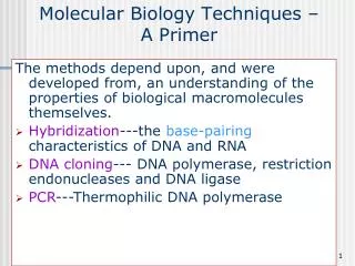
Molecular Biology Techniques. Nicky Mulder Acknowledgements: Anna Kramvis for lecture material (adapted here). Experiments for different cell processes. Two levels of experiment. Small-scale -1-10 genes/proteins: PCR Restriction enzymes Cloning Hybridization

E N D
Molecular Biology Techniques Nicky Mulder Acknowledgements: Anna Kramvis for lecture material (adapted here)
Two levels of experiment • Small-scale -1-10 genes/proteins: • PCR • Restriction enzymes • Cloning • Hybridization • Large-scale 100-10000 genes or whole genome -> High-throughput biology
Polymerase Chain Reaction Fnlmh.ufl.edu/cowries/PCR
Agarose gel electrophoresis • Agarose is used to form a gel • Gel is placed in solution with an anode and cathode • DNA has net negative charge from sugar-phosphate backbone –migrates towards anode • Migration speed is determined by size • Run DNA with some markers of known size • Visualized by ethidium bromide –flouresces in UV light
Restriction enzymes • Restriction enzymes recognize specific or defined 4 to 8 base pairsequences on DNA and cut
Restriction maps on gel - + www.cbs.dtu.dk
Use of restriction enzymes • Cloning • Restriction fragment length polymorphism
Restriction Fragment Length Polymorphism (RFLP) M1 M2 A2 A1 D con 1.5 kb 1.0 kb 750 bp 671 bp 593 bp 500 bp 448 bp 400 bp 300 bp 200 bp 145 bp uncut StuI
Cloning www.biodavidson.edu
Cloning vectors Features: Antibiotic resistance gene Another marker gene (lacZ*) Specific promoter Multiple cloning site *Lac Z gene, encodes beta-galactosidase- causes bacteria expressing the gene to appear blue when grown on a medium that containing X-gal
Other vectors • Bacterial artificial chromosomes • Yeast artificial chromosomes • Organism-specific vectors • Expression vectors
Prokaryote gene transfer • Conjugation –transfer between bacteria by direct contact • Transduction –transfer of DNA via a virus • Transformation –uptake of DNA from environment by competent cells
http://www.cdc.gov/ncidod/eid/vol6no1/images/vanderpoel1b.gifhttp://www.cdc.gov/ncidod/eid/vol6no1/images/vanderpoel1b.gif
Northern Hybridization http://www.molecularstation.com/images/northern-blot-med.jpg
Western Hybridization http://www.steve.gb.com/images/science/western_blotting.png
High-throughput biology • Move away from single gene focus and bottom-up approach • Studying multiple genes at once • Using new technologies • Moving from genotype to phenotype • Trying to find function of sets of genes: Functional genomics
Functional genomics experiments • DNA sequencing and analysis • Mutagenesis and gene disruption • DNA microarrays (transcriptomics) • Proteomics (protein expression, 2D gels, protein-protein interactions) • Structural genomics • Metabolomics
Functional genomics & Bioinformatics • Large-scale experiments generating vast amounts of data • Data needs sorting and analysis • Bioinformatics allows: • Tracking of samples • Automating data capture • Data storage and analysis • Data mining to convert data into biological research
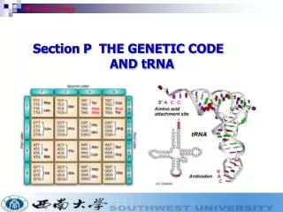
DNA sequencing technologies • Sanger sequencing method (chain termination) • Dideoxynucleotide triphosphates (ddG/A/T/C/TP, lack 3-OH), labelled primers and DNA polymerase -4 reactions –run on gel • Dye terminator sequencing • Label terminators with diff dyes –single reaction, use capillary electrophoresis • High-throughput sequencing • Parallel reactions, DNA on surfaces –sequencing by synthesis and detection of fluorescence
Sanger sequencing method www.bio.davidson.edu/Courses/Bio111/dnaseq2.gif
Genome sequencing • To sequence a fragment of DNA: • subclone fragment into vector- plasmid (2kb), cosmid (40kb), BAC (>100kb) or YAC (1Mb) • Grow cells and purify DNA • Sequence user flourescent dye labels and laser detection –can get 300-800bp per read • Problem is if fragment is too big –not covered by reads
Whole genome shotgun • Need to fragment the DNA, sequence the pieces and then assemble them • Need to over-sample to get good overlaps • May still get gaps using this approach, but can design new primers for additional sequencing • Repeats are an issue –can cause incorrect assembly • Shotgun sequencing works for small genomes like bacterial genomes
Sequencing complex genomes • As the complexity increases so does likelihood of incorrect assembly –eukaryotes has many repeats • Genome maps are important and form a guide for showing positions of genes and features • Eukaryotic genomes are fragmented into 1.5Mb bits and cloned into BACs, then a shotgun approach is used for each BAC –hierarchical shotgun sequencing • These “contigs” are assembled as before, and mapped onto genome using markers (genetic map)
Hierarchical shotgun sequencing International Human Genome Sequencing Consortium, 2001, Nature 409, pg 860-921.
Assembly Hierarchical shotgun sequencing PHRED Base calling, trace quality, Crossmatch –finds vector CONSED sequence editing PHRAP Assembly: Align fragments, consensus quality Highest quality reads used for consensus
Genome Annotation • Two main levels: • Structural Annotation – Finding genes and other biologically relevant sites thus building up a model of genome as objects with specific locations • Functional annotation – Objects are used in database searches (and expts) aim is attributing biologically relevant information to whole sequence and individual objects
Genome structure Gene prediction Promoter prediction Genes, pseudogenes, introns, exons, intergenic regions Proteins Translation BLAST Signatures 2D structure 3D structure Functional annotation
Annotation can be at different levels Function, structure Interactions, pathways Gene regulation Cellular process, localisation
Gene expression -Transcriptomics • Microarrays • ChIP on chip (Chromatin IP on microarrays)
Microarray overview Slide with target deposited label cDNA (probe) hybridise labelled probe to slide wash slides scan analyse results
Experimental design Image processing Normalization Pre-processing Data analysis Data mining Microarray data analysis
Data mining Add gene identifiers Add gene descriptions Add GO terms -AB02387 -SB07593 -AA00498 -AC008742 -AB083121 -GO0003456 -GO0006783 -GO0142291 -GO0054198 -GO0000234 -RNA polymerase -Glycosyl hydrolase -Phosphofructokinase -Transcription factor -Glucose transporter Map onto pathways
Proteomics • Large-scale study of proteins to determine their function • Proteome is protein complement of the genome • Includes the study of: • Protein structure and function • Protein-protein interactions • Protein expression • Protein localization • Protein modifications
Proteomics studies Mass spectrometry Xray, NMR Mass spectrometry Localization studies
Sample preparation Protein separation Protein selection Protein identification Workflow of a proteomics experiment Sample can be from patient cohort, cell selection, fraction, etc. Different separation techniques, e.g. 2-D PAGE, HPLC, ICAT, etc. Depends on separation method Usually mass spectrometry
Protein separation • 2D PAGE • Gel-free systems: • ICAT • HPLC Mass spec –digest proteins further
Protein separation -2D PAGE Size gradient pH gradient
Bioinformatics component • Sample tracking • Image capture • Image analysis and comparison: • Measuring intensities • Removing background noise • Finding difference between gels
Mass spectometry • Digest proteins with e.g. trypsin (lysine or arginine) • Proteins ionized and brought into gas phase • Move through mass analyzer which separates them based on mass • Detector records presence of ions
Protein identification (MS) Peptide Fragment Fingerprinting (PFF)
Peptide identification (MS/MS) VHLTPEEKSAVTALWGKVNVDEVGGEALGRLLVVYPWTQRFFESFGDLSTPDAVMGNPKVKAHGKKVLGAFSDGLAHLDNLKGTFATLSELHCDKLHVDPENFRLLGNVLVCVLAHHFGKEFTPPVQAAYQKVVAGVANALAHK Recognises lysine (K) & arginine (R) digest with trypsin denature V HLTPEEK VH LTPEEK VHL TPEEK VHLT PEEK VHLTP EEK VHLTPE EK VHLTPEE K Mass spec VHLTPEEK SAVTALWGK VNVDEVGGEALGR LLVVYPWTQR FFESFGDLSTPDAVMGNPK VK AHGK K VLGAFSDGLAHLDNLK GTFATLSELHCDK LHVDPENFR LLGNVLVCVLAHHFGK EFTPPVQAAYQK VVAGVANALAHK mass spectrum compare with theoretical peptide spectra; ID = best similarity