Download
1 / 23
250 likes | 746 Views
Coronary Artery Disease and Acute Myocardial Infarction. NPN 200 Fall 2006. Coronary Artery Disease. Atherosclerosis Define: thickness and hardening of the arteries caused by deposits of fat and fibrin which harden.

E N D
Coronary Artery Disease and Acute Myocardial Infarction NPN 200 Fall 2006
Coronary Artery Disease • Atherosclerosis • Define: thickness and hardening of the arteries caused by deposits of fat and fibrin which harden. • Leads to decreased lumen and decreased blood flow and ischemia and death of the tissue • Arteriosclerosis • Define: loss of elasticity and abnormal thickening or hardening of the walls of the arteries which can be due to accumulation of lipids, cholesterol, calcium or thrombus. • May also lead to occlusion of the lumen of the vessel, usually at the bifurcation of the vessels • May develop collateral circulation if develops slowly
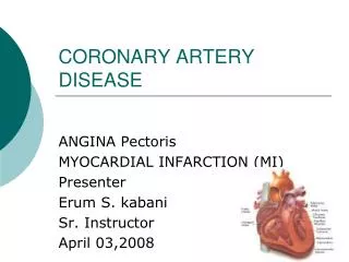
Signs and Symptoms • Usually none until > 60% • LAD most effected • Pain usual symptom but may experience dyspnea • May have irregular heart rate • N/V may also accompany the other symptoms • Called angina • Unstable – persistent, even at rest • Prinzmetal's – variant, and may occur without atherosclerosis
Medical Treatment • Decrease risk factors • Diet • Control cholesterol/triglycerides • Exercise • Smoking • Hypertension • Drugs • Calcium channel blockers • Nitroglycerin • Low dose ASA • Surgery
Myocardial Infarction • Myocardial infarction is the necrosis of an area of cardiac tissue as a result of obstruction of blood flow through a coronary artery or one of its branches • The myocardial tissue dies as a result of the occlusion • The size and location of the necrosed area affects the heart’s ability to squeeze • Death occurs from this cardiac damage or complications R/T to the MI • ½ of deaths occur within 1 hour after the onset of symptoms
Complications of an MI • Cardiogenic shock • Arrhythmias • CHF • Ventricular rupture or aneurysm • Pericarditis • Pulmonary embolism • Post-myocardial infarction
Risk Factors • Smoking • Family history • Hypertension • Elevated triglycerides and cholesterol levels • Obesity • Sedentary lifestyle • Aging • Stress • Men more than women (but women are increasing) • Diabetes mellitus
Causes • Arthrosclerosis (90%) • Constriction or spasm of the coronary artery • Coronary artery embolus • Coronary artery thrombus
Assessment for Chest Pain • Subjection • Tightness, heaviness, squeezing, or crushing pain in the substernal area, which can radiate to the jaw, neck, left arm, or shoulder • Determine if pain is precipitated by an event (exercise, stress or exertion) • Is the pain relieved by rest or drugs? • Is there any predisposing factors? • URI, PE, Hypoxemia, blood loss • Patient may experience anxiety and feeling of doom
Assessment for Chest Pain, cont. • Objective • Dyspnea • N/V • Profuse diaphoresis • Adventurous breath sounds • Tachycardia, decreased B/P, ^ temp • Elevation of cardiac enzymes (CPK, CPK-MB, AST, LDH, Troponin) • EKG changes • Results of any procedures completed
Medical Treatment • Early treatment is important • Goal is to preserve myocardial tissue • Nitroglycerin • Dilates coronary arteries • Morphine sulfate – 2-4 mg titrated for pain relief • decreases blood return to the heart • decreases anxiety • relaxes smooth muscle in the lungs • has analgesic effect
Medical Treatment, cont. • Oxygen at 2-4 L/min • Thrombolytic therapy – must meet criteria • Streptokinase • TPA • Heparin • ASA • Lidocaine, Calcium channel blockers, Digoxin, Beta blockers, Dopamine, Dobutamine • Angioplasty/Stent placement • Coronary Artery Bypass Grafting • Transmyocardial Lazer revascularization
Nursing Interventions For MI • Provide quiet, calm environment • Keep client on bedrest for 24-48 hours • Give medications as ordered –analgesics, O2, Nitroglycerin • Elevate head of bed • Watch for any more chest pain • Maintain IV line • Monitor for signs of CHF, cardiogenic shock, and pulmonary edema • Evaluate signs of MI • Skin color, and temperature • Monitor vitals • Observe EKG for dysrhythmias • Monitor fluid volume levels • Check labs
Continued Care of MI • Cardiac Rehab • Begin as soon as patient is stable • Individualized for need • Involves stages • Includes nurse, physician, nutritionist, physical therapy and social workers • Home care • Teach about medications • Include follow-up with physician • May need to teach about CAD • Teach modification of risk factors –weight, diet, smoking, exercise, etc. • Notify of any chest pain
Cardiopulmonary Arrest • Sudden cessation of hearts pumping function, stopping ventilation and circulation • Rapidly fatal if untreated • Accounts for > 350,000 deaths/year • Prompt treatment and early hospitalization necessary to prevent death • Causes • MI • V-Fib • Heart failure • Electrolyte imbalances • Hemorrhage • Electrical shock
Objective Symptoms • Unconscious • Absence of pulse and respirations • Absence of heart sounds • Pupillary dilation • Cyanosis
Diagnostic Tests • History • Physical • EKG • Enzymes after emergency treatment
Implementation • CPR • ABC’s • IV for administration of drugs • ABG’s frequently • Give Lidocaine, etc. • Watch for hypoxia, arrythmias, acidosis, and hypokalemia • Monitor labs • Assess LOC, skin color, temp, pulses, seizures, pupil changes • Observe for complications (rib fractures, tamponade, pneumothorax) • Give emotional support to the family