Download
1 / 39
400 likes | 630 Views
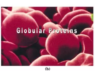
Globular Proteins. Figure 8-35 X-Ray diffraction photograph of a single crystal of sperm whale myoglobin. Page 240. Figure 8-39a Representations of the X-ray structure of sperm whale myoglobin. ( a ) The protein and its bound heme are drawn in stick form. Page 244.

E N D
Figure 8-35 X-Ray diffraction photograph of a single crystal of sperm whale myoglobin. Page 240
Figure 8-39a Representations of the X-ray structure of sperm whale myoglobin. (a) The protein and its bound heme are drawn in stick form. Page 244
Figure 8-39b Representations of the X-ray structure of sperm whale myoglobin. (b)A diagram in which the protein is represented by its computer-generated Ca backbone. Page 244
Figure 8-39c Representations of the X-ray structure of sperm whale myoglobin. (c)A computer-generated cartoon drawing in an orientation similar to that of Part b. Page 244
Figure 8-43a The H helix of sperm whale myoglobin. (a)A helical wheel representation in which the side chain positions about the a helix are projected down the helix axis onto a plane. Page 247
Cut-away view surface Stryer Fig. 3.45 Mb yellow = hydrophobic, blue=charged, white=others
Structural features of most globular proteins: 1. Very compact: e.g. Mb has room for only4 water molecules in its interior. 2. Most polar/charged R groups are on the surface and are hydrated. 3. Nearly all the hydrophobic R groups are on the interior. 4. Pro occurs at bends/loops/random structures and in sheets
Chapter 9!!! Figure 9-1 Page 277
Figure 9-2 Reductive denaturation and oxidative renaturation of RNase A. Page 277
Figure 9-3 Plausible mechanism for the thiol- or enzyme-catalyzed disulfide interchange reaction in a protein. Page 278
Figure 9-14b Reactions catalyzed by protein disulfide isomerase (PDI). (b) The oxidized PDI-dependent synthesis of disulfide bonds in proteins. Page 288
Figure 9-4 Primary structure of porcine proinsulin. Page 278
H-bond Fun Fact • 1984 survey of protein crystal data shows that “almost all groups capable of forming H-bonds do so.” (main chain amides, polar side chains)
Many conformational states Fewer conformational states A “single” conformational state
High energy Many conformational states Fewer conformational states A “single” conformational state Low energy
Figure 9-11c Folding funnels. (c) Classic folding landscape. Page 285
Figure 9-11d Folding funnels. (d) Rugged energy surface. Page 285
“Ideal” “Real” ?
Figure 9-12 Polypeptide backbone and disulfide bonds of native BPTI. Page 286
Figure 9-13 Renaturation of BPTI. Page 287
Figure 9-28 Conformational fluctuations in myoglobin. Page 303
Figure 9-30a The internal motions of myoglobin as determined by a molecular dynamics simulation. (a) The Ca backbone and the heme group. Page 305
Figure 9-30b The internal motions of myoglobin as determined by a molecular dynamics simulation. (b) An a helix. Page 305
Figure 9-32a Amyloid fibrils. (a) An electron micrograph of amyloid fibrils of the protein PrP 27-30. Page 307
Figure 9-32bc Amyloid fibrils. (b) and (c) Model and isolated b sheet. Page 307
Figure 9-34a Evidence that the scrapie agent is a protein.(a) Scrapie agent is inactivated by treatment with diethylpyrocarbonate, which reacts with His side chains. Page 310
Figure 9-34b Evidence that the scrapie agent is a protein.(b) Scrapie agent is unaffected by treatment with hydroxylamine, which reacts with cystosine residues. Page 310
Figure 9-34c Evidence that the scrapie agent is a protein.(c) Hydroxylamine rescues diethylpyrocarbonate-inactivated scrapie reagent. Page 310
Figure 9-35a Prion protein conformations. (a) The NMR structure of human prion protein (PrPC). Page 311
Figure 9-35b Prion protein conformations. (b) A plausible model for the structure of PrPSc.
Figure 9-36 Molecular formula for iron-protoporphyrin IX (heme). Page 313
Figure 9-37 Primary structures of some representative c-type cytochromes. Page 313
Figure 9-38 Three-dimensional structures of the c-type cytochromes whose primary structures are displayed in Fig. 9-37. Page 314