Download
1 / 16
180 likes | 333 Views
Immunopatholgy RSV blood flukes. The diseases in which pathology is largely caused by the immune response, is known as immunopathology

E N D
The diseases in which pathology is largely caused by the immune response, is known as immunopathology • This is true to some degree in most infections; for example, the fever that accompanies a bacterial infection is caused by the release of cytokines by macrophages • Most important examples are • Leprosy √ • respiratory syncytialvirus (RSV) infection • Blood flukes
In adults, it may only produce symptoms of a common cold, such as a stuffy or runny nose, sore throat, mild headache, cough, fever, and a general feeling of being ill • But in premature babies and kids with diseases that affect the lungs, heart, or immune system, RSV infections can lead to other more serious illnesses
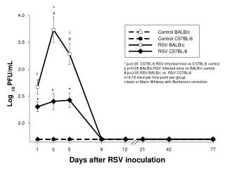
respiratory syncytial virus (RSV) infection • Bronchiolitis caused by RSV is the major cause of admission of young children to hospital • 4500 deaths out of 90,000 cases each year in the US alone • The first indication that the immune response to the virus might have a role in the pathogenesis of this disease came from the observation that infants vaccinated with an alumprecipitated killed virus preparation had a more severe illness than children who did not receive the vaccine • This occurred because the vaccine failed to induce neutralizing antibodies but succeeded in producing effectorTH2 cells • When the vaccinated children encountered the virus, the TH2 cells released interleukins IL-3, IL-4, and IL-5, which induced bronchospasm, increased the secretion of mucus, and increased tissue eosinophilia
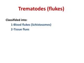
Another example of a pathogenic immune response is the response to the eggs of schistosomes (blood flukes) • These helminth parasites lay their eggs in the hepatic portal vein • Some reach the intestine and are shed in the feces, spreading the infection • other eggs lodge in the portal circulation of the liver, where they elicit a potent immune response leading to chronic inflammation, hepatic fibrosis, and eventually liver failure • This process reflects the excessive activation of TH1 cells and can be modulated by TH2 cells, IL-4, or CD8 T cells, which can also produce IL-4 TH2 TH1
Adaptive immune responses elicited by potentially harmless antigens i.e. not associated with infectious agents leading to harmful (immune mediated) hypersensitivity reactions known generally as allergic reactions • e.g. response to inherently harmless 'environmental‘ antigens such as pollen, food, and drugs
Anaphylaxis- History • The ability of the immune system to respond inappropriately to antigenic challenge was recognized early in this century • Two French scientists, Paul Portier and Charles Richet, investigated the problem of bathers in the Mediterranean reacting violently to the stings of Portuguese Man of War jellyfish • They knew that the localized reaction of the bathers was the result of toxins • Their first attempt met with disastrous results. • Portierand Richet injected dogs with the purified toxins, followed later by a booster of toxins • Instead of producing antibodies against the toxins, the dogs immediately reacted with vomiting, diarrhea, asphyxia, and, in some instances, death • Clearly this was an instance where the animals “overreacted”to the antigen • Portierand Richet coined the term anaphylaxis, loosely translated from Greek to mean the opposite of prophylaxis, to describe this overreaction • Richet - awarded the Nobel Prize in Physiology or Medicine in 1913 • Phylaxis- Protection against infection
Classification of types of hypersensitivity reactions by Coombs and Gell • Hypersensitivity reactions due to immunological responses were classified into four broad types by Coombs and Gell • Hypersensitivity reactions due to immunological responses were classified into four broad types according to • Type of immune response I.e. humoral or cell mediated • antigens • Types I-III are antibody-mediated and are distinguished by the different types of antigen recognized and the different classes of antibody involved • Type I is IgE • while types II and Ill are mediated by lgG • Type IV is cell- mediated responses
Type I hypersensitivity • Type I hypersensitivity reactions in this classification are immediate-type allergic reactions mediated by IgE antibodies • but many of the allergic diseases that are initiated by IgE antibodies, such as allergic asthma, have chronic features characteristic of other types of immune response, particularly of TH2 cell-mediated type IV hypersensitivity • Classical allergic reactions are dependent upon previous exposure and sensitization to specific allergens that result in the development of antigen specific IgEantibodies • Classical allergic reactions occur when allergens bind and crosslink allergen specific IgE antibodies present on the cell membranes of mast cell and basophils, triggering the release of numerous me
Allergic symptoms – due to inflammatory mediators In hay fever (allergic rhinoconjunctivitis), for example, symptoms occur when allergenic proteins leached out of grass pollen grains come into contact with the mucous membrane of the nose and eyes A predisposition to become IgE-sensitized to environmental allergens is called atopyboth genetic and environmental- that may contribute to predisposition
Fig. lgE-mediated reactions to extrinsic antigens. All lgE-mediated responses involve mast-cell degranulation, but the symptoms experienced by the patient can be very different depending, for example, on whether the allergen is injected directly into the bloodstream, is eaten, or comes into contact with the mucosa of the respiratory tract
IgE • IgE may protect external mucosal surfaces by promoting inflammation, enabling IgG, complement proteins, and leucocytes to enter the tissues • The main biological role of IgEis thought to be in adaptive immunity to infection with parasitic worms (helminths) and arthropods • The Fc portion of IgEmade against parasitic worms and arthropods can bind to eosinophils enabling opsonization • It is often made in response to allergens • IgEmakes up about 0.002% of the serum antibodies with a half-life of 2 days • Most IgE is tightly bound to basophils and mast cells via its Fcregion • IgE is a monomer and has 2 epitope-binding sites
T he first signal (indicated as 1 in the figure) required for B-cell activation is delivered through its antigen receptor (top panel). For thymus-dependent antigens- the second signal (indicated as 2) is delivered by a helper T cell that recognizes degraded fragments of the antigen as peptides bound to MHC class II molecules on the B-cell surface (center panel); the interaction between CD40 ligand (CD40L, also called CD154) on the T cell and CD40 on the B cell contributes an essential part of this second signal. For thymus-independent antigens, a second signal can be delivered along with the antigen itself, through Toll like receptors that recognize antigen associated TLR ligands, such as bacterial lipopolysaccharide (LPS) or bacterial DNA (bottom panel)
Fig. Helper T cells stimulate the proliferation and then the differentiation of antigen-binding • B cells. • The specific interaction of an antigen-binding B cell with a helper T cell leads to the expression of the B -cell stimulatory molecule CD40 ligandon the helper T-cell surface and to the secretion by the T cell of the B cell stimulatory cytokines IL-4, IL-5, and IL-6, which drive the proliferation and differentiation of the Bcell into • antibody-secreting plasma cells • memory cell