Download

1 / 60
630 likes | 1.45k Views
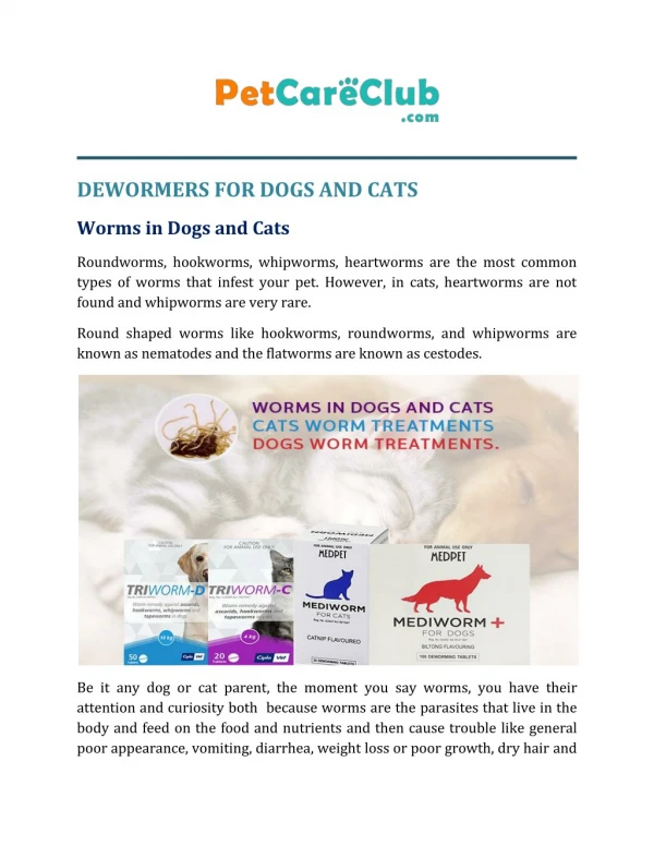
Ascarids of Dogs and Cats Toxocara canis Toxocara canis (common dog ascarid) is the most important of the two canine ascarids from the standpoint of prevalence, virulence and public health importance.

E N D
Ascarids of Dogs and Cats
Toxocara canis Toxocara canis (common dog ascarid) is the most important of the two canine ascarids from the standpoint of prevalence, virulence and public health importance.
T. canis occurs in about 15-20% of dogs of all ages, except in sparsely populated, dry areas where infection rates are less (arrow refers to incidence in Hawaii). Puppies have much higher incidence rates however, because of maternally acquired prenatal and perinatal infections. Without evidence to the contrary, it is often presumed that virtually all puppies are infected with larvae of T. canis.
Toxocara canis adults in petri dish Toxocara canis is a typical ascarid in that it is large and fairly robust. The females commonly are 12-15 cm. long by about 2 mm. wide. Males are somewhat shorter and thinner. When found in the ingesta of a freshly dead dog, they usually are coiled tightly, but after the carcass has cooled, the parasites relax and straighten. The anterior end of both sexes is bowed ventrally. The name, Toxocara, means "bowed head”.
T . canis anterior end This slide shows two major features of Toxocara canis. One is the 3 large, fleshy lips typical of all ascarids. These lips are used for "browsing" on internal epithelial surfaces - they are not suited for attachment. The other feature is the 2 wing-like cervical alae typical of T. canis. These alae are cuticular inflations which arise from the lateral body wall and may be seen grossly. They occur in both sexes at the bowed portion of the anterior end.
T. canis - The Swimmer Ascarids do not attach to the mucosa but rely on maintaining their position in the gut by a powerful swimming ability .
Clinical effects of T. canis are greatest in puppies. A thorough knowledge of the life cycle is necessary for a complete understanding of the pathogenicity of Toxocara canis. Effects are primarily due to adult worms, although migrating larvae are sometimes directly responsible for virulence.
There are four possible ways dogs can become infected by T. canis: • egg ingestion • transplacental transfer • transmammary transfer • ingestion of paratenic (transport) hosts
Toxocara canis, like most ascarids, are prolific egg producers and may produce > 100,000 eggs/day/female worm. Eggs become infective after 10-15 days on the ground at normal temperatures and contain an infective second stage larvae protected by a thick highly resistant egg wall (for other nematode groups, third stage larvae are infective). Infective eggs may remain viable for years. They survive cold well but are killed at temperatures above 37°C. After ingestion of infective eggs by dogs, the infection may take one or two courses depending on age, sex and previous exposure of the host.
After ingestion by pups less than 6 months of age, and especially in pups less than 5 weeks of age, the L2 is released in the gut, migrates via the hepatic-tracheal route, and returns to the digestive tract. Second stage larvae reach the liver via the hepatic portal system in 1-3 days, the lungs as third stage larvae at 3-5 days, the stomach as L4 by 10 days, and are in the duodenum as young adults (L5) by 3-4 weeks. The prepatent period for infection via the hepatic-tracheal route is 4-5 weeks.
Somatic Migration Both an age resistance and an acquired resistance due to a previous exposure occurs against T. canis infections. Suckling or weanling pups are fully susceptible, but become progressively more resistant thereafter until age resistance reaches maximal levels at about 6 months of age. True acquired immunity would constitute an additive resistance factor. As resistance builds, more and more larvae migrating through the lungs cross over into the somatic circulation and are disseminated into various tissues where they are trapped in small granulomas as dormant second stage larvae. These “arrested” larvae remain viable for years.
Transplacental(prenatal) infection When a bitch becomes pregnant, latent somatic larvae are activated at 30-40 days of gestation and larvae migrate across the placenta into the liver and lungs of the fetus during the last trimester. This is thought to be the major life cycle survival strategy and is the main way to insure infection levels in dog populations .
In the fetus, juvenile larvae accumulate in the liver and lungs until whelping. At birth they resume migration by the hepatic-tracheal route and most larvae (L5) are in the small intestine by the 6th day of life. Infection by the prenatal route results in a more abbreviated prepatent period than that resulting from direct ingestion of eggs by pups, and eggs are passed as early as 2-3 weeks of age. Although most larvae migrate to the fetus in one pregnancy, three pregnancies are required to completely clear the tissues of a bitch, assuming there is no further exposure to Toxocara canis eggs.
Transmammary Passage “Activated" larvae have also been shown to accumulate in the mammary glands to be later acquired by suckling pups . Larvae have been found in milk taken at birth and up to 22 days after parturition. Larvae in the milk assume a typical hepatic-tracheal migration.The importance of this mode of infection is thought to be minor (2% ) and the transplacental route is most important for Toxocara canis in dogs, accounting for 98% of infections in pups. It is commonly observed that an increase in Toxocara infections occurs in nursing bitches. There are two possible explanations for this: 1) infection of a bitch may occur by passage of partially mature larvae in the feces of pups, and subsequent ingestion during cleaning of pups; larvae pass to the small intestine and mature directly without further migration, or 2) some " activated " larvae may find their way to the gut of the bitch and mature there. The effect in either case is that the bitch and her pups may harbor ascarids of exactly the same stage of maturation.
Ingestion of paratenic hosts Although it is a minor source of infection it is possible for dogs to become infected by ingestion of a variety of paratenic hosts (including rats, mice, swine, sheep, birds, and others) which contain arrested L2 stages in somatic tissues. After ingestion, direct development occurs without hepatic-tracheal migration
Damage due to Toxocara canis consists primarily of digestive disturbances due to worms in the intestinal tract, but there may be lesser signs due to migrating forms, especially in the liver and lungs.
Liver lesions Hemorrhagic tracts and foci occur in the liver due to migrating larvae a few days after infection. eosinophilic, granulomatous cellular infiltrates result histologically and rapid healing occurs with little or no contribution to clinical signs of ascariasis .
T. canis Lung Pathology In the lungs, usual infections result in small petechiae or ecchymoses when larvae break out of capillaries into alveoli.
T. canis Lung Pathology In very heavy infections, large numbers of larvae simultaneously passing through the lungs may lead to lobular pneumonia with the alveoli filled with edema, RBC's, eosinophilic exudate and larvae. Lesions progress to granulomatous interstitial change. Lung signs, if they occur,are seen 3-7 days post-infection in association with massive infections which on very rare occasions may be lethal. Very heavy maternal infections of pups can result in acute death of entire litters at 2-3 weeks of age due to pulmonary sequelae of massive Toxocara migration +/- dehydration associated with diarrhea and vomiting.
Adult worms in duodenum Presence of adult worms in the intestine result in irritation and mild catarrhal enteris manifested as diarrhea and sometimes vomiting. Direct effects ascribed include competition with the host for nutrients, damage from ascarid "browsing" on villi and an obscure interference with normal mechanisms of the host absorptive process. Intestinal obstruction is very unusual even with very large numbers of worms. Blockage due to migration up bile ducts may occur on rare occasions. Adult worms live an average of 4 months in the proximal small intestine.
Intestinal epithelium-histosection Microscopically, lesions consist of relatively mild catarrhal inflammation with an increase in the number of goblet cells and mild mucosal infiltration with plasma cells, lymphocytes and eosinophils.
Eosinophilia A variable eosinophilia in peripheral blood is seen with heavy chronic infections. Eosinophilia is classically seen clinically as a result of infection by helminths, some ectoparasites, and in allergic conditions.
Clinical Signs Toxocara canis infections may result in the unthriftiness, dull hair coat and the "poor-doer" pups that are typical of chronic parasitism. Pups are stunted and have a "pot-bellied” appearance. Digestive disturbances include vomiting, diarrhea and constipation. Worms are often presented to veterinarians for identification after being found in vomitus by owners. Young pups may die of weakness and dehydration if heavy infections are not treated. Some clinicians can detect a "sweetish odor" to the breath.
T. canis in Older Dogs Older dogs are more resistant to T. canis and clinical signs, when they occur, are generally manifested only as unthriftiness, or diarrhea. The resistance of older dogs may be explained on the basis of both age resistance and acquired resistance due to prior exposure.
Age resistance. A true age resistance of obscure mechanisms begins at weaning and becomes progressively stronger until it reaches a maximum at about six months of age. The percentage of animals shedding T. canis eggs in the feces of mature (over 1 year) and immature dogs (0-6 months) over a five year period at Iowa State University is presented in this chart. The percentage of infected immature dogs was 40-50% (white bars) versus an infection rate of 5-10% in mature dogs (red bars).
Self-cure The acquisition of resistance results in an incompletely understood phenomenon called" self -cure”, which occurs with other nematodes in addition to Toxocara canis. A local anaphylactic reaction in the digestive tract is postulated as the mechanism of self cure and results in the clinical observation of diarrhea, high fever (pyrexia) and passage of ascarids in feces.
Mechanisms of self-cure On the basis of experimental evidence, it is theorized that ascarid antigens combine with local mast cells at surface IgE receptors. At a critical Ag-Ab level, the mast cells are "triggered" to lyse, producing histamine-like substances which in turn cause release of locally produced secretary IgA antibody and increased mucosal permeability. Mucosal infiltration with inflammatory cells occurs concurrently. The net effect of this "local anaphylaxis" reaction is the damage of worms by products of inflammation, antibody and/or cells from the mucosa and expulsion of the worms. The old practitioner's adage that "a fever causes the passage of ascarids" may be due to this phenomenon.
Juvenile ascarid: in vitro antibody In vitro studies on the immune response to ascarids provide support for the self-cure theory . This Toxocara juvenile has been incubated in the serum of an ascarid-infected dog. Casts and plugs have formed from antigen-antibody reactions around the juvenile's mouth. Such reactions may weaken the worm and impede its activity, but will not kill the juvenile outright.
Mechanism of larva encapsulation A second manifestation of immunity against ascarid juveniles is the in vitro attraction and adhesion of leukocytes to the juveniles cuticular integument (as seen here). The counterpart of this reaction in tissues would act on larvae migrating through tissues and explain their encapsulation.
Granulomatous foci due to ascarid encapsulation in the kidney Large numbers of larvae are "arrested" in dormancy or are killed in tissues of resistant hosts (as seen grossly in this kidney). Granulomatous encapsulation of larvae is typical in resistant hosts and is believed to be a cell-mediated or delayed hypersensitivity type response.
Diagnosis of Toxocara canis Diagnosis of Toxocara canis infection may be done by demonstrating typical large, dark, thick-shelled eggs at fecal examination. Identification of worms in vomitus or diarrhea is also diagnostic.
Toxascaris leonina Toxascaris leonina has a wide distribution in the U.S. but has a much lower incidence as compared to Toxocara in the south. (Some call it the "northern roundworm").
Toxascaris leoninaHosts Toxascaris leonina occurs in dogs, cats and various wild canidae and felidae.
Toxascaris leonina adult male and female. Toxascaris is morphologically similar to Toxocara. Females measure 10 cm. and males are up to 7 cm long.
Cervical alae Cervical alae and other major morphological characters are similar in appearance to Toxocara canis and these two species are difficult to differentiate on this basis.
Toxascaris eggs The eggs of Toxascaris leonina are easily differentiated from either Toxocara species, and can be found by fecal examination or by dissecting mature eggs from adult female specimens .
Mucosal Development The life cycle of Toxascaris leonina is much simpler than Toxocara.Toxascaris leonina does not normally utilize the hepatic-tracheal or somatic migration patterns and prenatal infections do not occur. Infections occur by direct ingestion of eggs or via transport (paratenic) hosts. After ingestion of infective eggs (eggs are infective after about 1-2 weeks on the ground) larval development occurs in the wall of the small intestine. Second stage larvae enter into the intestinal wall for 9-10 days and return directly to the lumen where they molt twice to produce fourth stage larvae (L4) by 35 days, molt to L5 (sexually immature adult stage) by 6 weeks and produce eggs at about 75 days post-infection. Toxascaris is therefore not found by fecal examination until 2 1/2-3 months of age at the earliest (vs. 2-3 weeks for T. canis).
Paratenic hosts. When mice and other small abnormal hosts ingest infective eggs, larvae hatch out and migrate to various body tissues and become encapsulated. Dogs, and especially cats, may become infected by ingestion of these paratenic hosts .
Clinical effects of Toxascaris leonina are similar to Toxocara in the digestive tract (mild catarrhal enteritis) but since larvae do not migrate, there is no liver and lung involvement. The potential for infection is not as high as for Toxocara since prenatal or transmammary does not occur, and infections in puppies under 2 1/2-3 months of age are not seen.
Toxascaris vs. Toxocara eggs Demonstration of typical eggs in feces is diagnostic or adults may be identified in vomitus, feces or at necropsy. Toxascaris leonina (right side) eggs are more oval, slightly smaller and lighter in color than Toxocara (left side) and the shell is "rough on the inside" with some space observed between the embryo and shell as opposed to Toxocara eggs which are "roughened on the outside" with a very dark, complete filling of the shell.
Toxocara cati T. cati is morphologically similar to Toxascaris leonina, which also occurs in cats, but may be differentiated by the broader "arrowhead" cephalic alae and the "bowed" head.
T. cati egg Adult worms live in the small intestine of cats and produce eggs (similar in morphology to T. canis) that become infective 3-4 weeks after passed. When eggs are ingested, larvae undergo hepatic-tracheal migration as L2's which return to the GI tract, enter the stomach wall for a time, return to the lumen and after 3 molts are found in the small intestine as L5's. The prepatent period is longer than T. canis, about 8 weeks .
Transport hosts-mouse A number of animals can serve as paratenic hosts of T cati, especially mice and rats. Since migration occurs in paratenic hosts, no migration is seen in the cat and direct development to adults occurs in the gut.
Transmammary transmission Transmammary passage of larvae occurs in colostrum and throughout the first three weeks of lactation. This mode of transmission is the most important source of infection in kittens. Transplacental (prenatal) infections are not thought to occur. Gut lesions are similar, but other damage due to T. cati is less severe than T. canis for several possible reasons. Infections obtained by transmammary or transport hosts undergo direct development in the gut and do not migrate by the hepatic-tracheal route like T .canis. Although a migratory phase occurs after ingestion of infective T. cati eggs, the effect of larvae on lungs is less than T. canis, possibly because no molts occur in the liver and lungs .
Toxocara cati Clinical Signs Clinical signs occur mostly in young animals and consist of stunting, pot-belly, diarrhea, poor coat and occasional deaths related to dehydration and weakness in heavy infections. Diagnosis is made by finding typical eggs or identifying adults. Heavily infected kittens are common and often show dramatic improvement in general health and condition following treatment. Remember: weanling cats may not yet be shedding eggs.
Treatment of Ascarids of Dogs and Cats A wide assortment of drugs are available for use against ascarids. Older drugs are often cheap but have narrower spectrum against other parasites and include: Piperazine (ascarids only), methylbenzene (®Methycide) and dichlorvos (®TASK, ®TASK-tabs). Newer drugs include the broad spectrum benzimidazoles (Mebendazole, Fenbendazole, Febantel, Oxybendazole) and the avermectin's (®Ivomec, milbemycin) but are generally more expensive. Parantel pamoate (®Nemex) has gained wide routine usage in puppies and kittens because of its safety and easy acceptability of the malt flavored liquid vehicle. Parantel has only moderate effectiveness (80-90%) against mature ascarids and hookworms, however, and 2 or more follow-up treatments 1-2 weeks apart are often required to completely clear infections. Fenbendazole (SID for 3 days) has the advantage of high efficacy against both adult and immature L4 stages.
Treatment of Ascarids of Dogs and Cats Diethylcarbamazine (®DEC) is reasonably effective (80%) in preventing new ascarid infections in concentrations used for prophylaxis of heartworms. The addition of oxybendazole to DEC (®filarabits-plus) produces a highly effective preventive against ascarids, hookworms and whipworms. Milbemycin (®Interceptor) is effective against roundworms, hookworms, and whipworms at the heartworm preventive dose. The addition of pyrantel to ivermectin (®Heartgard-Plus) adds efficacy against both roundworms and hookworms. Preventive heartworm medications that have efficacy against roundworms are effective and can be used in lieu of this regimen if they are begun at the recommended time at weaning .
Prevention and Sanitation If used with preventative sanitation measures listed below, drugs are currently available to allow near eradication of ascarids in both kennel and household environments. A good rule is to treat pups at 2,4, and 6 weeks after parturition and at vaccinations thereafter. Kittens (and queens) should be treated at 4 and 6 weeks of age (prior to egg shedding by worms) and at vaccinations . This will prevent the passage of eggs by the pup or kitten. Repeat treatment at 2-4 week intervals is needed to get adults produced from new infections or larvae which may have been in tissues at time ofthe first treatment. Ascarid burdens are known to increase in nursing bitches for unclear reasons. Bitches should be treated before parturition toreduce egg exposure of pups and again on the same schedule used for the litter (at 2 weeks and each 2-4 weeks thereafter).
Prevention and Sanitation Since eggs require an incubation period of 2 weeks or more outside the host before they become infective, cleaning of cages at frequent intervals and disposing of feces containing eggs help lower the contamination by eggs. Use of 10% chlorox makes eggs less "sticky", allowing hoses to be effective. Construct kennel so that all of the floor is exposed to direct sunlight sometime during the day (at least 2 hours). While the eggs of T. canis are resistant to desiccation and chemicals, they are killed by sunlight, UV light, and high temperature. Feed and water resources should be high enough to avoid fecal contamination.