Download
1 / 73
740 likes | 1.05k Views
The Immune System. Chapter 15. Objectives of Chapter 15. To understand the: Major lines of immune defense Mechanisms of action of B and T lymphocytes Active and passive immunity, vaccines Immunological tolerance. Defense Mechanisms. Against pathogens constitute the immune system

E N D
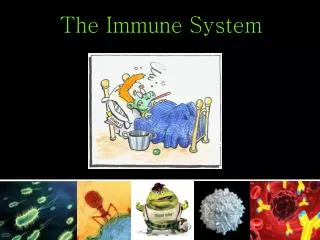
The Immune System Chapter 15
Objectives of Chapter 15 • To understand the: • Major lines of immune defense • Mechanisms of action of B and T lymphocytes • Active and passive immunity, vaccines • Immunological tolerance
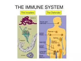
Defense Mechanisms • Against pathogens constitute the immune system • Grouped into 2 categories: • Innate (nonspecific) immunity is inherited as part of structure of each organism • Adaptive (specific) immunity is a function of lymphocytes and changes with exposure
Innate Immunity • Distinguishes between “self” and “non-self” • by recognizing molecules of PAMPs (pathogen-associated molecular patterns) • First line of defense against invading pathogens • Includes epithelial barriers, high acidity of gastric juice, phagocytosis, interferons, and fever
Activation of Innate Immunity • Triggered in response to PAMPs produced only by microorganisms • Best known are lipopolysaccharide (LPS) from gram negative bacteria and peptidoglycan from gram positive bacteria • Some immune cells have receptors for PAMPs displayed on their surfaces • Receptors called Toll receptors • Ten different Toll receptors identified in humans
Phagocytosis • Performed by 3 classes of phagocytic cells: • Neutrophils: first to arrive at infection sites • Mononuclear phagocytes: macrophages and monocytes • Organ-specific phagocytesin liver, spleen, lymph nodes, lungs, and brain • Fixed phagocytesline sinusoids of liver, spleen, and lymph nodes and remove pathogens
Phagocytosis Pseudopods from phagocyte surround pathogen forming a vacuole • Pseudopods fuse forming a complete vacuole, lysosomal enzymes restricted to the organelle • Lysosome fuses with vacuole febore complete fusion of pseudopods is complete, lysosmal enzymes released into the infected area of tissue
Fever • Appears to be component of innate immunity • Occurs when hypothalamic thermostat is reset upwards by interleukin-1 and other cytokines (endogenous pyrogens) • Pyrogens released by WBCs in response to endotoxin from gram negative bacteria • Interleukin-1, interleukin-6, tumor necrosis factor • Cytokines also increase sleepiness and a fall in the plasma Fe2+ concentration • functions in inhibiting bacterial activity
Interferons • Polypeptides produced by cells infected with virus • provide short-acting, non-specific resistance to viral infection in nearby cells • 3 types – alpha, beta, and gamma interferon • Most body cells make alpha and beta interferons • act to protect other cells near viral infection • Viruses able to penetrate these cells but replication is inhibited • Gamma interferon porduced only by specific lymphocytes and natural killer cells
Life Cycle of HIV • RNA virus – injects RNA into host cell • Viral RNA transcribed by reverse transcriptase into complementary DNA (cDNA) • Enters nucleus integrates into host DNA • cDNA directs synthesis of RNA that 3) Codes for viral proteins that assemble new viral particles 4) New viruses budded from host cell to infect other cells
Adaptive Immunity • Acquired ability to defend against specific pathogens • by prior exposure to those pathogens • Mediated by production of specific antibodies by lymphocytes
Antigens • Molecules that elicit a response • production of antibodies that specifically bind to the antigens • Usually large molecules that are foreign to the body • Immune system can distinguish “self” molecules from non-self antigens • Normally makes antibodies only against non-self antigens • Most naturally occuring antigens have many antigenic determinant sites • different sites stimulate production of different antibodies with specificities for these sites
Haptens • Small non-antigenic molecules – become antigens when bound to proteins • form an antigenic determinant site • Useful for creating antibodies for research and diagnosis • Bonding of foreign haptens to a person’s proteins can occur • Derivatives of penicillin can produce fatal allergic reactions in susceptible people
Immunoassays • Tests that use specific antibodies to identify a particular antigen • Binding of antibody to antigen causes clumping (agglutination) • which can be visualized
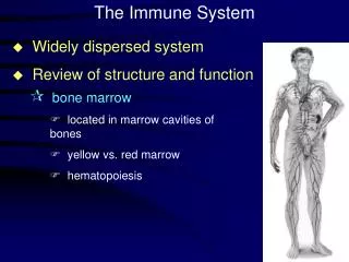
Lymphocytes • Derived from stem cells in bone marrow • Which replace selves by cell division so are not depleted • Lymphocytes produced by this process seed thymus, spleen, and lymph nodes with self-replacing colonies
T Lymphocytes (T cells) • Develop from lymphocytes that seed thymus • Do not secrete antibodies • Attack infected host cells, cancer cells, and foreign cells • Thus provide cell-mediated immunity • Supply 65 – 85% of lymphocytes for the blood • and most of lymphocytes in germinal centers of lymph nodes and spleen
B Lymphocytes (B cells) • Become immunocompetent in bone marrow • Fight bacterial infections by secreting antibodies into blood and lymph • Thus provide humoral immunity
Thymus • Located below the thyroid gland • Grows during childhood, gradually regresses after puberty • Contains T cells that supply other tissues • T cells can be depleted, e.g. by AIDs or chemotherapy • These can only be replenished up to late childhood • After that, repopulation is accomplished by production in secondary lymphoid organs
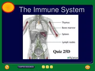
Secondary Lymphoid Organs • Consist of lymph nodes, spleen, tonsils, and Peyer’s patches • Located in areas where antigens could gain entry to blood or lymph • Lymphocytes migrate constantly through blood and lymph from one lymphoid organ to another • Enhances chance that antibody will encounter its antigen • Spleen filters blood; other lymphoid organs filter lymph
Local Inflammation • Occurs when bacteria enter a break in the skin • Inflammatory reaction – initiated by nonspecific mechanisms, phagocytosis and complement activation • Complement activation attracts phagocytes to area
Local Inflammation • As inflammation progresses, B cells produce antibodies against bacterial antigens • Attachment of antibodies to antigens amplifies nonspecific responses because of complement activation • And promotes opsonization:ability of antibodies to promote phagocytic activity (neutrophils, macrophages, monocytes)
Events in a Local Inflammation Bacterial antigens • bind to antibodies, which coat the bacteria – this activates complement and • Promotes phagocytosis by neutrophils ¯ophages – activation of complement also • Stimulates mast cells – release histamine and other mediators of inflammation thhat promote capillary permeability and • Extravasation of leukocytes to inflamed site
Local Inflammation • In inflamed area, leukocytes attach to surface of endothelial cells • Move by chemotaxis to inflamed site • Neutrophils arrive first, then monocytes, then T cells • Undergo extravasation 15-26
Local Inflammation • Mast cellssecrete heparin, histamine, prostaglandins, leukotrienes, cytokines, and TNF-a • produce redness, warmth, swelling, pus, and pain • Recruit more leukocytes • If infection continues, endogenous pyrogens are released
B Lymphocytes (B cells) • Have antibodies on surface that are receptors for antigens • When bound to antigen, are stimulated to divide and secrete antibodies
B Lymphocytes • When B cells divide, some progeny become memory cells • Others become plasma cells produce about 2000 antibodies/sec • Specific for original antigen • This provides active immunity
B Lymphocytes (B cells) • Binding of B cells to antigen also triggers a cascade of reactions that activate complement proteins • Complement proteins can kill antigen-bearing cells and promote phagocytosis
Antibodies • Proteins called immunoglobulins • gamma globulinclass of plasma proteins • Antibodies have same basic structure but their differences provide for antibody specificity
Antibody Structure All antibodies consists of four polypeptide chains • Shape of “Y” • 2 long heavy (H) chains joined to 2 shorter light (L) chains • When cleaved, stalk of Y becomes crystallizable fragment (Fc) • Fc is constant among different antibodies • Arms of Y contain antigen-binding fragment (Fab) • Fab contains a variable region that confers antibody specifity
Diversity of Antibodies • Each person has about 1020 antibody molecules • With a few million different specificities • Likely there is an antibody specific for any antigen a person might encounter • 2 mechanisms might account for diversity • If a few hundred genes code for Heavy chains and a few hundred for Light chains, different combinations could lead to millions of different antibodies • Recombination of these in developing lymphocytes of marrow produces antigen-independent diversity
Antibody Diversity • Diversity further increases via somatic hypermutation • in which there is a high rate of single base pair mutations • Occurs as B cells undergo proliferation in 2o lymphoid tissues in response to foreign antigens • Antigen-dependent diversification also occurs in genetic recombination • A switch in constant regions of heavy chains of antibodies so that IgM are converted to IgA or IgE (class switch recombination)
The Complement System • Part of nonspecific defense system • Activity triggered by binding of antibodies to antigens (classic pathway) and by • unique polysaccharide coating of bacteria (alternative pathway) • Binding of antibodies to antigens does not by itself destroy antigens or pathogens • Antibodies label targets for complement system attack and also • stimulate opsonization (ability of antibodies to stimulate phagocytosis)
The Complement System • Series of proteins whose activation forms a membrane attack complex • perforates a cell membrane causing it to undergo lysis • Complement proteins can be subdivided into 3 functional groups: • C1: recognition • C4, C2, C3: activation • C5-C9: attack (complement fixation) • These form the membrane attack complex
The Complement System • Classic pathway – antibody-antigen binding activates C1 • C1 hydrolyzes C4 into C4a and C4b • C4b binds to cell membrane and becomes active, splitting C2 into C2a and C2b • C2a attaches to C4b and cleaves C3 into C3a and C3b • Alternate pathway also cleaves C3 (and so converges with classic pathway at this step) • C3b converts C5 to C5a and C5b
Membrane Attack Complex • C5b and C6 - C9 are inserted into bacterial cell membrane, forming membrane attack complex • creates large pore in membrane, causing osmotic influx of H2O, lysis, and cell death
Complement Fragments • Liberated during activation have a number of effects, including: • Attracting phagocytes (chemotaxis) • Phagocytes have receptors for C3b which can serve as bridge to victim cell (opsonization) • C3a and C5a stimulate mast cells to release histamine • Which increases blood flow and capillary permeability, bringing in more phagocytes
Killer or Cytotoxic T Cells • Carry CD8 cell surface marker • Destroy body cells that possess foreign antigens • Usually from a pathogen, but can be from malignancy or self cells never seen by immune system • Kill by cell-mediated destruction • That is, must be in contact with victim cell • Kill by secreting perforins which create a pore in victim's membrane and cause lysis • Also secrete granzymeswhich cause destruction of victim's DNA
Helper and Suppressor T Cells • Helper T-cells carry CD4 surface marker • Indirectly participate by enhancing responses of both killer T-cells and B- cells • Regulatory T-cellsdecrease responses of killer T-cells and B cells • Carry CD25 surface marker (and CD4) • Help protect against autoimmune responses
Lymphokines • Cytokines secreted by lymphocytes • Usually called interleukin-1, 2, 3 (or IL-1, IL-2 )
T Cell Receptor Proteins • Only protein antigens are recognized by most T cells • T cell receptors cannot bind to free antigens • T cells respond to foreign antigens when they are presented on surface of antigen-presenting cells • Chief antigen presenting cells are macrophages and dendritic cells
Dendritic Cells • Originate in marrow, then migrate to most tissues • Prominent where pathogens might enter body • Engulf protein antigens, partially digest them, and display polypeptide fragments on surface for T cells to "see“ • Fragments associated on surface with histocompatibility antigens which are necessary to activate T-cells
Dendritic Cells • To increase chance of interacting with correct T-cells, dendritic cells migrate to secondary lymphoid organs • secrete chemokines to attract T-cells
Histocompatibility Antigens • On surface of all body's cells except mature RBCs • Also called human leukocyte antigens (HLAs) • Coded for by group of 4 genes on chromosome 6 called the major histocompatibility complex (MHC) • The 4 genes have multiple alleles creating many possible MHC types
MHC • MHC genes produce 2 types of cell surface molecules: class-1 and class-2 • Class-1s are made by all cells except RBCs • Class-2s are made only by antigen-presenting cells and B-cells • These present class-2s, together with foreign antigens, to helper T-cells • This is the only way to activate helper T-cells so they can promote B-cell activity
MHC • In order for killer and helper T-cells to function they require co-presentation of antigen with a specific MHC marker • Killer T-cells are activated to kill victim cell only by co-presentation of antigen and class-1 marker • Helper T-cells require antigen and class-2 marker
Interactions Between Antigen Presenting Cells and Lymphocytes • Co-presentation requirement comes from presence of different coreceptors on killer and helper T cells • Killer T coreceptor CD8 interacts only with class-1s • Helper T coreceptor CD4 interacts only with class-2s
T Cell Response to a Virus • When virus infects the body it is phagocytized by macrophage or dendritic cell • Partially-digested polypeptide fragments are antigens displayed on cell surface • Form a complex with class-2 MHC molecules that macrophages present to helper T-cells • Helper T-cells bind and are activated • Can now promote B-cell activity