Download

1 / 20
250 likes | 1.1k Views
Detecting Pelvic Disease With Duplex Ultrasound. Ron Bush, MD, FACS Midwest Vein & Laser Center Dayton, Ohio. Why is a Scan Being Performed?. Varicose veins Swelling Possibility of DVT Venous claudication Pelvic symptoms. Consider.

E N D
Detecting Pelvic Disease WithDuplex Ultrasound Ron Bush, MD, FACS Midwest Vein & Laser Center Dayton, Ohio
Why is a Scan Being Performed? • Varicose veins • Swelling • Possibility of DVT • Venous claudication • Pelvic symptoms
Consider • An ultrasound (US) of the pelvis includes an US of the femoral vein • Abnormal femoral US occurs frequently in pelvic compression or obstruction • Phasic flow is the normal pattern on doppler US
Ultrasound Findings: Cardiac • Back and forth pulsatile flow (roller coaster) on US may indicate: • ASD • Tricuspid regurgitation • AV fistula
Ultrasound Findings: Obesity • Alteration in normal flow pattern • May be evidence of reflux at femoral & popliteal level • Usually symmetric • Often overlooked as cause of venous hypertension of the extremities
Ultrasound Findings: Acute Thrombus • No flow signal or monophasic pattern may be present • Non-compressibility of the iliac and/or femoral vein • Acute symptoms usually present
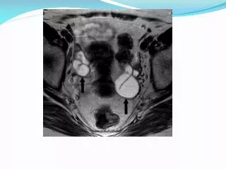
Nutcracker Syndrome • Left renal vein compression between the aorta and superior mesenteric artery • Can lead to pelvic congestion syndrome • May be associated with greater saphenous vein (GSV) insufficiency through collaterals • May present with unusual pattern of varices on thigh or leg
Gluteal Varicosities (Bush Venous Lectures, 2011)
Ultrasound of LRV Compression (Bush Venous Lectures, 2011)
Ultrasound Findings: Nutcracker Syndrome • Stenosis evaluated by comparing the anterior posterior (AP) diameter of the left renal vein (LRV) on left side of the aorta and at level of stenosis • Ratio of AP diameter and ratio of peak velocities should be used as US criteria for stenosis
Nutcracker Syndrome: Diagnosis With Doppler US. (Kim, 1996)
May-Thurner Syndrome • Compression of left iliac vein by right common artery • May be associated with leg swelling, varicosities, or iliac vein thrombosis with resultant sequalae
Unilateral Leg Swelling (Bush Venous Lectures, 2011)
Ultrasonic Diagnosis of Iliac Vein Compression (May-Thurner) Syndrome (Levent, 2007)
Ultrasound Findings: May-Thurner Syndrome • Evidence of compression • Monophasic flow may be present distal to obstruction • High velocity flow at area of stenosis
Ovarian Vein Reflux • Manifestations may Include • Pelvic congestive syndrome • Vulvar varices • Inguinal or thigh varices • Ovarian vein > 4mm
Vulvar Vein (Bush Venous Lectures, 2011)
Conclusion Any of the following should alert you to altered flow patterns in the pelvic veins: • Varicosities in unusual locations • Unilateral leg swelling • Acute thrombosis of the iliac vein • Reflux or monophasic flow on femoral US • Vulvar varices
References Thombosis femoral vein image. Retrieved May 7, 2011 online from http://www.answers.com/topic/deep-vein-thrombosis. Nutcracker image. Retrieved May 7, 2011 online from www.phlebolymphology.org/2009/07/nutcracker-syndrome/ Kim SH, Cho SW, Kim HD, Chung JW, Park JH, Han MC. Nutcracker syndrome: diagnosis with Doppler US. Radiology. 1996;198:93-97. Leg image. Retrieved May 7, 2011 online from http://www.bushvenouslectures.com/blog/content.asp?id=348Oguzkurt L, Ozkan U, Tercan F, Koc Z, Sadick N, Trelies, M. A clinical histological and computer-based assessment of the Polaris LV, combination diode, and radio frequency system for leg treatment. Diagn Interv Radiol 2007;13:152-155.