Download
1 / 63
840 likes | 2.78k Views
Myelodysplastic Syndrome. Ahmad Sh. Silmi Msc Haematology, FIBMS. What Is Myelodysplastic Syndrome?. The myelodysplastic syndromes are a group of disorders characterized by one or more peripheral blood cytopenias secondary to bone marrow dysfunction .

E N D
Myelodysplastic Syndrome Ahmad Sh. Silmi Msc Haematology, FIBMS
What Is Myelodysplastic Syndrome? • The myelodysplastic syndromes are a group of disorders characterized by one or more peripheral blood cytopenias secondary to bone marrow dysfunction. • In MDS the bone marrow cannot produce blood cells effectively, and many of the blood cells formed are defective. • These abnormal blood cells are usually destroyed before they leave the bone marrow or shortly after entering the bloodstream. • As a result, patients have shortages of blood cells, which are reflected in their low blood
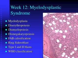
Characteristics • Varying degree of tri-lineage cytopenia ( red blood cells, white blood cells and platelets). • Dysplasia • Normocellular or hypercellular B.M. • May progress to acute leukaemia
Signs and Symptoms • Excessive tiredness, shortness of breath, and pale skin can be caused by anemia (shortage of red blood cells). • Serious infections with high fevers can be caused by leukopenia(not having enough normal white blood cells) and, in particular, by having neutropenia or granulocytopenia(too few mature granulocytes). • Excessive bruising and bleeding, for example, frequent or severe nosebleeds and/or bleeding from the gums, can be due to thrombocytopenia (not having enough of the blood platelets needed for plugging holes in damaged blood vessels).
Incidence 1- Disease of elderly. 2- Median age is 65 years. 3- <10% are younger than 50 years. 4- Incidence rates 1/100,000 pop./ years. 5- Incidence rise to 1/1000 / years in > 60 years old. 6- Male slightly higher than female
MDS Etiology • Two etiologic categories of MDS: 1.) De Novo: Associated with: -benzene exposure (gasoline) -cigarette smoking -viruses -Fanconi’s anemia 2.) Therapy related: Associated with: -alkylating agent chemotherapy -radiation
Aetiology This shows thetow arms of haemopoiesis
Aetiological Agents • Tobacco smoke. • Ionizing radiation. • Organic chemicals (such as benzene, toluene, xylene, and chloramphenicol). • Heavy metals. • Herbicides. • Pesticides. • Fertilizers. • Stone and cereal dusts. • Exhaust gases. • Nitro-organic explosives. • Petroleum and diesel derivatives. • Alkylating agents. • Marrow-damaging agents used in cancer chemotherapy.
Chromosomal abnormalities and MDS • Chromosomal abnormalities are present in up to 50%of de novo cases of MDS and in virtually all cases of secondary MDS. The most common are: • Abnormality - 7 +7 +8 5q- 7q- 11q- 12q- 13q- 20q- inv3 i(17q) t(1;3) t(1;7) t(3;3) • Frequency(%) 15 5 19 27 4 7 5 2 5 1 5 1 2 1
Deletion of the long arm of chromosome 5 (5q- syndrome ) • Strongly associated with RA. • 5q- accounts for up to 70% of cytogenetic abnormalities in this subtype. • The q arm of chromosome 5 is particularly rich in genes, which encoded haemopoietic growth factors and their receptors.For example , IL-3 , IL-4 , IL-5 , GM-CSF and the M-CSF receptor are located in this region. • The potential for the loss of any or all of these genes contribute to the disruption of ordered haemopoiesis.
Monosmy 7 and 7q- • Most strongly associated with secondary MDS. • Associated with the loss of a major surface glycoprotein (gp 130) in neutrophile and susceptibility to bacterial infection secondary to impaired granulocyte monocyte chemotatic activity.
Deletion of the q arm of chromosome 11 (11q-) • Account for 20% of the chromosomal abnormalities in RAS. • This abnormality is associated with raised iron stores and high ring sidroblast counts. • The presence of the gene , which encoded the H-subunit of ferritin at chromosome 11 , may explain this
Abnormalities of chromosome 17 (i17q) • It involves the loss or disruption of the Р53 tumor suppressor gene are seen in CML in association with transformation to the blastic phase and in up to 5% of cases of primary MDS. • This predisposes to certain dysplastic features and neutrophil vaculation.
Abnormalities of chromosome 3 • Dysmegakaryopiesis and thombocytosis appear to be associated with Abnormalities of chromosome 3
The importance of indication of chromosomal abnormalities • To confirm diagnoses . • To know the stage of disease. • To know the direction of progression of disease. • Multiple genetic abnormalities indicate late events in MDS.
Patterns of MDS evolution • A stable group with no increase in B.M blasts and a normal karyotype . • Rapid blast transformation with acquisition of new cytogenetic changes after an initial , stable phase . • Gradually increasing blast count in the absence of new cytogenetic changes .
Refractory Anemia • RA Definition: • Dyplasia of the erythroid series only. • Clinically, anemia is refractory to hematinic therapy • Myeloblasts < 1% blood and < 5% marrow • <15% ringed sideroblasts in marrow • No Auer rods • Other etiologies of erythroid abnormalities must be excluded. These include: • drug/toxin exposure -vitamin deficiency • viral infection -congenital disease
Refractory Anemia • Epidemiology: • 5-10% of MDS cases. • Older patients • Morphology: • Anisopoikilocytosis on peripheral smears • Dyserythropoiesis with nuclear abnormalities (megaloblastoid change) • < 15% ringed sideroblasts
Refractory Anemia • Genetics: • 25% may have genetic abnormalities • Prognosis: • Median survival is 66 months • 6% rate of progression to acute leukemia
Refractory Anemia with Ringed Sideroblasts • RARS definition: • Dyplasia of the erythroid series only. • Clinically, anemia is refractory to hematinic therapy • Myeloblasts < 5% in marrow, absent in blood • >15% ringed sideroblasts in marrow • No Auer rods • Other etiologies of ringed sideroblasts must be excluded. These include: • Anti- tuberculosis drugs • Alcoholism
Refractory Anemia with Ringed Sideroblasts • Epidemiology: • 10-12% of MDS cases. • Older patients • Males > females • Morphology: • Dimorphic pattern on peripheral smears • Majority RBC’s normochromic, 2nd population hypochromic • Dyserythropoiesis with nuclear abnormalities (megaloblastoid change)
Refractory Anemia with Ringed Sideroblasts • Morphology (con’t.) • < 15% ringed sideroblasts (RS) • RS = Erythroid precursor with ≥ 10 siderotic granules encircling 1/3 or more of the nucleus. • If excess blasts present, this dictates diagnosis, despite percentage of RS’s.
Refractory Anemia with Ringed Sideroblasts • Genetics: • Clonal chromosomal abnormalities in • <10%; in fact, development of such an abnormality should prompt reassessment of diagnosis. • Prognosis: • Median survival 6 years (72 months) • 1-2% rate of progression to acute leukemia
Refractory Anemia with Excess Blasts • RAEB definition: • Refractory anemia with 5-19% myeloblasts in the bone marrow. • RAEB-1: • 5-9% blasts in bone marrow and <5% blasts in blood. • RAEB-2: • 10-19% blasts in the bone marrow • Auer rods present
Refractory Anemia with Excess Blasts • Epidemiology: 40% of MDS cases. • Older patients (over 50 years) Morphology: • Dysplasia of all three cell lines often present • Neutrophil abnormalities may include: • Hypogranulation -hypersegmentation • Pseudo-Pelger-huet (hyposegmentation/barbells) • Pseudo Chediak-Higashi granules • Megkaryocyte abnormalities may include • Hypolobation -Micromegakaryocytes
Refractory Anemia with Excess Blasts • Morphology (con’t.) • Erythroid precursor abnormalities may include: • Abnormal lobulation -megaloblastoid change • Multinucleation • 0-19% myeloblasts in the blood • 5-19% in the marrow • Bone marrow: • Usually hypercellular (10-15% hypocellular) • Abnormal localization of immature precursors (ALIP) may be present • Immunophenotype: • Blasts express CD 13, CD33 or CD117 • The only MDS with a relevant phenotype
Refractory Anemia with Excess Blasts • Genetics: • Clonal chromosomal abnormalities found in 30% - 50% of RAEB cases. The abnormalities include: • +8 – -5 – del(5q) – -7 – del(7q) – Complex karyotypes • Prognosis: • Median survival, RAEB-1 = 18 months • Median survival, RAEB-2 = 10 months • RAEB-1 = 25% rate of progression to acute leukemia • RAEB-2 = 33% rate of progression to acute leukemia
Chediak-Higashi-like Granules • Photograph courtesy of John Scariano, University of New Mexico, Dept. of Pathology
Refractory Anemia with Excess Blasts in Transformation (RAEB-t) • 21-30 percent blasts in the marrow; more than 5 percent in the bloodstream • excessive numbers of monocytes (a type of white blood cell) • normal or hypercellular (filled with cells) marrow • accounts for about 25 percent of cases
Chronic Myelomonocytic Leukemia (CMMOL) • 5-20 percent blasts in the marrow; less than 5 percent in the bloodstream • cytopenia of at least two cell lines • normal or hypercellular (filled with cells) marrow • accounts for 15 to 20 percent of cases.
CMML • Splenomegaly (10%) • Maculopapular skin infiltration • Monocytic pleural or pericardial effusion • JMML (MPD/MDS) • Pallor, bleeding, hepatosplenomegaly, skin involvement
WHO • Refractory anemia • Refractory anemia e ringed siderblast • Refractory cytopenia e multilineage dysplasia • Refractory cytopenia e multilineage dysplasia & ringed sideroblasts • Refractory anemia e excess blast-1 • Refractory anemia e excess blast-2 • Myelodysplastic syndrome unclassified • MDS associated e isolated del (5q)
Prognostic Groups • Two groups based on survival and evolution to acute leukemia • 1.) “Good” group • Refractory anemia (RA) • Refractory anemia with ringed sideroblasts (RARS) • 5q - syndrome • 2.) “Bad” group • Refractory anemia with excess blasts (RAEB) • Refractory anemia with excess blasts in transformation (RAEB-t) • CMML • MDS unclassified can be either
International Prognostic Scoring System (IPSS) Factors • the percentage of blasts in the bone marrow. • whether chromosome abnormalities are present and, if so, which ones. • how low the patient's blood counts are. These are given a score; the lowest scores have the best outlook for survival.