Download
1 / 48
500 likes | 962 Views
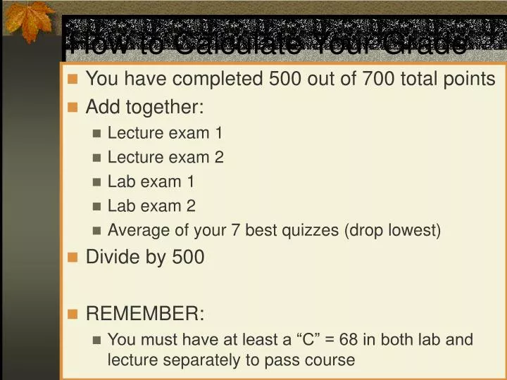
How to Calculate Your Grade. You have completed 500 out of 700 total points Add together: Lecture exam 1 Lecture exam 2 Lab exam 1 Lab exam 2 Average of your 7 best quizzes (drop lowest) Divide by 500 REMEMBER:

E N D
How to Calculate Your Grade • You have completed 500 out of 700 total points • Add together: • Lecture exam 1 • Lecture exam 2 • Lab exam 1 • Lab exam 2 • Average of your 7 best quizzes (drop lowest) • Divide by 500 • REMEMBER: • You must have at least a “C” = 68 in both lab and lecture separately to pass course
How to Calculate the Number of points you need to pass • You need a total of 476 out of 700 points to get a C • Take the total number of points you just calculated (the sum of 4 exams and quiz average) and subtract it from 476 • The number you have is the total number of points you need • If you divide that number by 2, you will see the approximate grade you’ll need on lab exam 3 and lecture exam 3. • REMEMBER: the rules from previous page apply • Have to have at least C in lecture and lab separately
Organs of the Abdomen Systems: Urinary and Digestive
Urinary System • Kidneys • Purify blood • Ureters • Drain urine from kidney to bladder • Urinary Bladder • Store urine • Urethra • Drain urine from bladder to outside body pg 5
Kidneys: major excretory organs • Remove toxins, metabolic waste, excess H2O, ions • Urea, uric acid, creatinin • Regulates volume + makeup of blood • Maintains balance between • Salts and water • Acids and bases
Kidneys: Gross Anatomy • Located superior lumbar region • Posterior abdominal wall (T12-L3) • Retroperitoneal • Hilus • Adrenal Gland: superomedial to kidney • Renal Artery + Vein • Innervation: branches of renal plexus pg 648
Kidneys: Gross Anatomy • Renal Capsule • Layer of tough CT • Maintains shape • Prevents spread of infection • Adipose Capsule • External to renal cap • Perirenal fat • Surrounded by fascia • Keeps in place, cushions • Pararenal Fat • External to adipose cap • Keeps in place, cushions pg 649
Kidney: Internal Anatomy • Cortex • Superficial • Light, granular • Part of functional unit • Medulla • Deep layer • Darker • Pyramid-cone shape • Contain collecting tubule collect urine Pg 650
Kidney: Internal Anatomy • Medullary Pyramid • Base: against cortex • Apex: inward • Papilla = tip • Drips urine into minor calyx • Minor Calyx (calices) • Cup-shaped divisions of major calices • Surround papilla of pyramid • Major Calyx (calices) • Larger cup-shaped branches of renal pelvis • Renal Pelvis • Flat expansion of ureter • Collects urine Pg 650
Kidney: Microscopic Anatomy • Functional Unit • Uriniferous Tubule • Nephron • Collecting tubule • Waste is filtered out • Waste products formed • Located in lobes of kidneys pg 652
Ureters • Slender tubes transport urine • Run from kidneys to bladder • Retroperitoneal • Continuation of renal pelvis • Enters bladder at oblique angle to prevent backflow • Increased pressure in bladder closes distal end of ureter pg 648
Ureters: 3 Layers • External: Adventitia • CT • Middle: Muscularis • Smooth Muscle • Inner Longitudinal • Outer Circular • External longitudinal (on distal third) • Peristalsis • Inner: Mucosa • Transitional epithelium
Bladder • Muscular sac store and expel urine • Location • On pelvic floor • Posterior • Pubic symphysis • Anterior • Males = rectum • Females = vagina, uterus • Collapses + Expands • Full into abdominal cav • Emptystays in pelvic cav • Supplied by branches of internal iliac arteries + veins • Innervated = branches of hypogastric plexus pg 648
Bladder: Internal Anatomy • 3 Layers • Mucosa = transitional epithelium & lamina propria • Detrusor Muscle: smooth muscle • Inner/Outer longitudinal, Middle circular • Fibrous Adventitia = CT • Parietal peritoneum on superior surface instead trigone pg 662
Urethra • Drains urine from bladder to outside • Female = short tube • Males = long tube • Prostatic, Membranous, Spongy (penile) portions • Also carries semen • Internal Urethral Sphincter • Between bladder + urethra • Thickening of detrusor (smooth muscle) • External Urethral Sphincter • Within urogenital diaphragm • Skeletal muscle = voluntary control urination • External Urethral Orifice • Males = end of penile urethra • Females = anterior to vaginal opening, posterior to clitoris
Urethra: Female vs. Male pg 662
Micturition = Urination • Emptying bladder • Stretch receptors in bladder respond when bladder full • Parasympathetic signals detrusor muscle to contract and internal urinary sphincter to open (also inhibits sympathetic pathways that would prevent urination) • Other brain receptors can inhibit urination by relaxing detrusor, and keep external urinary sphincter closed • Voluntary contraction of abdominal wall muscles increases abdominal pressure • Voluntary relaxation of external urethral sphincter See pg 663
Digestion System • Alimentary Canal • Mouth • Pharynx • Esophagus • Stomach • Small Intestine • Large Intestine • Accessory Organs • Teeth, Tongue • Salivary Glands • Gallbladder • Liver • Pancreas pg 5
Food Processing Activities • Ingestion: taking food into mouth • Propulsion: food moves through gut • Swallowing + Peristalsis • Mechanical Digestion: breakdown of food • Chewing, Churning, Segmentation • Chemical Digestion: chemical breakdown • Enzymes • Absorption: Digestive end products into blood • Defecation: Removal of waste products
Alimentary Canal Wall • Internal = Mucosa + Submucosa • Epithelium • Lamina propria: • contains MALT: mucosa-associated lymphoid tissue • Muscularis mucosae • Submucosa = CT w/elastic fibers, nerves, vessels • Middle = Muscularis Externa • Inner circular layer • Outer longitudinal layer • Creates sphincters • Outer = Serosa or Adventitia
Innervation of Alimentary Canal • 2 Plexuses: Myenteric & Submucosal • Parasympathetic, Sympathetic, Visceral Sensory fibers • Enteric Nervous System • 100 million neurons in walls of alimentary canal = internal system • Within above plexuses • Independent reflex arcs • Controls glandular secretion, peristalsis, segmentation • Autonomic Nervous System speeds up or slows activity controlled by enteric system
Stomach • “J” shape • Cardiac Region • Junction esophagus • Cardiac sphincter (Gastroesophageal) • Fundus (“dome”) • Under diaphragm • Body • Large, middle part • Pylorus • Distal portion • Pyloric sphincter • Greater Curvature • Lesser Curvature Pg 624
Internal Anatomy of Stomach • Mucosa • Rugae: mucosal folds allow expansion • Many intrinsic glands • Goblet cells • Gastric glands • Typical Submucosa • Muscularis externa • Oblique layer • Circular layer • Pyloric sphincter • Longitudinal layer • Serosa pg 624
Function of Stomach • Temporary storage of chyme • Breakdown begins • Churn, segmentation • Pepsin proteins • Absorption • H2O, electrolytes • Alcohol, other drugs • Stays about 4 hours • Hold from1.5-4 liters
Small Intestine: Parts + Functions • Parts • Duodenum = proximal (5%) • Jejunum = middle (~40%) • Ileum = distal (~55%) • Majority of enzymatic digestion • Bile: emulsifier (gallbladder, liver) • Enzymes (pancreas) • Almost all nutrient absorption • Segmentation • Moves chyme around to increase contact with intestine walls • Food takes about 3-6 hours to move through • 2.7- 6 meters
Small Intestine: Internal Features • Intestinal flora: produce vitamin K • Simple columnar epithelium w/many modifications for absorption • Lymph tissue in submucosa • Muscularis externa has 2 layers • Some parasympathetic innervation from vagus • Arterial supply: • Superior mesenteric • Rt (cranial) pancreaticoduodenal
Small Intestine:Modifications of epithelium for absorption • Length • Increase surface area • Plicae circularis • Transverse ridges of mucosa • Increase surface area • Slow movement of chyme • Villi • Move chyme, increase contact • Contain lacteals: remove fat • Microvilli: • Increase surface area • Modifications decrease distally pg 629
Small Intestine • Duodenum: • short, straight • Mostly retroperitoneal • Jejunum & Ileum: • highly coiled • Fewer modifications • Hang by mesentery in peritoneal cavity • Mesentery Arcades • Arteries + veins • Nerves • Store fat Pg 614
Large Intestine • Cecum • Vermiform appendix • Colon • Ascending • Transverse • Descending • Sigmoid • Rectum • Anal Canal pg 631
Large Intestine • Functions: • Absorb water and electrolytes • Form, store and expel feces from body • Internal Features: • Intestinal flora • No intestinal villi or modifications for absorption • Many goblet cells • Simple columnar epithelium except lower half of anal canal • Significant Lymph tissue in mucosa & submucosa • Muscularis mucosae has 2 layers • Some parasympathetic innervation from vagus
Colon: External Features • Taeniae coli • 3 longitudinal strips • thickening of longitudinal muscle • maintain muscle tone • create haustra • Haustra • saclike divisions • Epiploic Appendages • fat-filled pouches • significance unknown pg 631
Cecum + Vermiform Appendix • Cecum • sac-like, blind pouch • Ileocecal valve • raised edges of mucosa • prevents feces going back into ileum • Vermiform Appendix • same layers • blind tube opens into cecum • masses of lymph tissue pg 631
Colon • Ascending colon • Right side • Hepatic flexure (= right colic flexure) • Transverse colon • Across cavity • Descending colon • Left side • Splenic flexure (= left colic flexure ) • Sigmoid colon • Enters pelvis • “S” shape pg 631
Colon: Function • Absorb H2O and electrolytes • Some digestion by bacteria • Mass Peristaltic Movements (2-3x day) • Moves through in 12-24 hours • 1.5 meters
Rectum + Anal Canal • Rectum • descends into pelvis • no teniae coli • longitudinal muscle layer complete • rectal valves • Anal Canal • passes through levator ani muscle • releases mucus to lubricate feces • Internal anal sphincter • involuntary, smooth m. • External anal sphincter • voluntary, skeletal m. • Stratified squamosal epithelium at lower half pg 632
Defecation Reflex • Stretching of rectum wall initiates reflex • Spinal cord - parasympathetic signals sigmoid colon + rectum to contract + anal sphincter to relax (involuntary) • If not ready-reflex ends- rectum relaxes • Reflex initiated again until you go! • Contraction of abdominal muscles, levator ani + diaphragm assists defecation (voluntary)
pg 610 Liver • Largest gland (3 lbs) • Location • Upper Right Quadrant • Mostly under ribcage • Highly vascular • Some functions • produce bile • pick up glucose • detoxify poison, drugs • make blood proteins • many others pg 635
Liver: External Features • Diaphragmatic surface • Right lobe (larger) • Left lobe • Falciform ligament • Fissure between • Visceral surface • Quadrate lobe • Caudate lobe • Both part of left lobe pg 635
Liver: Visceral Surface • Hepatic Vein (into inferior vena cava) • Porta Hepatis • Hepatic Artery (from abdominal aorta ) • Hepatic Portal Vein • Carries nutrient-rich blood from stomach + intestines to liver • Portal system = 2 capillary beds! • Hepatic Ducts (carry bile) pg 636
Gallbladder • Muscular sac • Between right + quadrate liver lobes • Bile is stored + concentrated • Bile: breaks down fats = emulsification • Bile • Produced by liver • Stored in gallbladder pg 610
Gallbladder continued • Mucosa & lamina propria • Simple columnar epithelium • Expandable mucosal folds • Smooth muscle layer • Thick connective tissue • Covered by serosa in places
Bile Ducts • Cystic duct • carries bile from gallbladder • Hepatic duct • carries bile from liver • Common Bile duct • joins cystic and hepatic • carries bile into duodenum pg 628
Movement of Bile • Bile secreted by liver continuously • Hepatopancreatic (Vater) ampulla • common bile + main pancreatic duct meet and enter duodenum • Sphincter of Oddi around it • closed when bile not needed for digestion • Bile then backs up into gallbladder via cystic duct • When needed gallbladder contracts, sphincters open pg 628
Pancreas • Retroperitoneal • Gland • Exocrine • digestive enzymes • Endocrine • hormone insulin • hormone glucagon • Location • curve of duodenum • extends to spleen pg 639
Ducts of Pancreas • Main Pancreatic duct • joins common bile duct • enters duodenum • Hepatopancreatic (Vater) ampulla • Accessory Pancreatic duct • enters duodenum in other location pg 628
Spleen • Largest lymph organ • Highly vascular • Function • remove blood-borne antigens (immune) • remove and destroy old/damaged blood cells • stores blood platelets • In fetus: site of hematopoiesis pg 639
Arterial Blood Supply to Abdominal Viscera • All branches of Abdominal Aorta • Anastomoses • Left + Middle colic • Left + Right gastric • Left + Right gastroepiploic • Cranial + Caudal pancreaticoduodenal • Deep Iliac Circumflex + Adrenolumbar • STUDY HAND OUT! MUST KNOW WHAT SUPPLIES WHAT!!
Hepato = liver Pancreatico = pancreas Cystic = gallbladder Gastro = stomach Splenic = spleen Adreno = adrenal gl Lumbar = lumbar region Epiploic = membrane-covered Mesenteric = mesentery Duodenal = duodenum Ileo = ileum Colic = colon Rectal = rectum Names give hints!