Download
1 / 89
950 likes | 1.9k Views
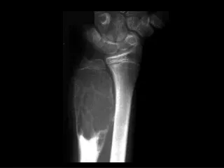
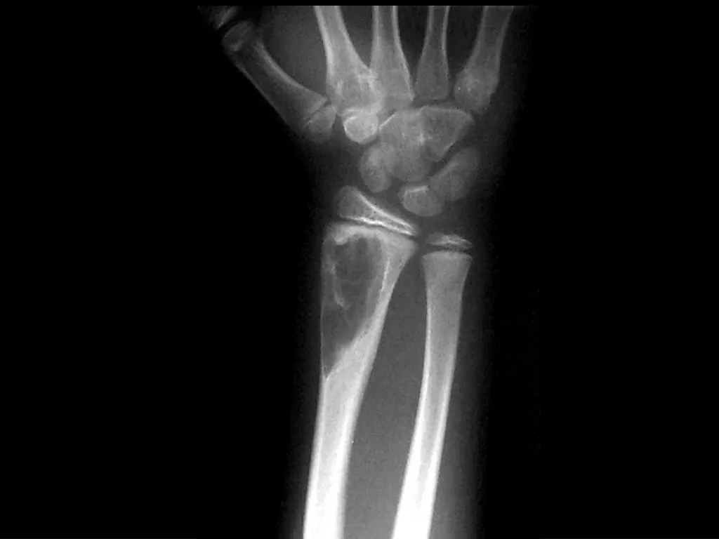
Aneurysmal bone cyst. Findings: Eccentric metadiaphyseal lucent lesion with a thin sclerotic margin and fine internal septa ddx: Fibrous dysplasia Chondromyxoid fibroma GCT, chondroblastoma (if physis closed). Adamantinoma. Findings:

E N D
Aneurysmal bone cyst • Findings: • Eccentric metadiaphyseal lucent lesion with a thin sclerotic margin and fine internal septa • ddx: • Fibrous dysplasia • Chondromyxoid fibroma • GCT, chondroblastoma (if physis closed)
Adamantinoma • Findings: • Expansile mixed lytic and sclerotic lesion of the tibial midshaft • Cortical disruption and periosteal reaction • ddx: • Fibrous dysplasia
Avulsion injury • Findings: • Soft tissue calcification along the superior anterior acetabulum • No cortical involvement • Normal joint space • ddx: • Myositis ossificans • Parosteal osteosarcoma
Brodie’s abscess • Findings: • Well-defined lytic lesion in the metaphysis • Large zone of surrounding sclerosis • CT is diagnostic of a fluid filled lesion • ddx: • Osteoblastoma • Langerhan’s cell histiocytosis
Calcaneal Ewing’s sarcoma • Findings: • Permeative lesion in the calcaneous • Cortical disruption and slight periosteal reaction • ddx: • Lymphoma • Metastasis/myeloma • Infection • Langerhan’s cell histiocytosis
Chondromyxoid fibroma • Findings: • Well-defined eccentric lucent lesion • Cortical thinning • Narrow zone of transition • ddx: • ABC • Infection • Fibrous dysplasia • Giant cell tumor (if physis closed) • Non-ossifying fibroma (if sclerotic margin)
Cortical desmoid • Findings: • Irregular cortical thickening of the posterior (medial) distal femur • Avulsion injury related to the medial gastrocnemius • ddx: • Parosteal osteosarcoma • Myositis ossificans
Langerhan’s cell histiocytosis • Findings: • Predominantly lytic lesion of the tibia with cortical thickening, periosteal reaction, and soft tissue swelling and edema • ddx: • Infection • Ewing’s sarcoma • Lymphoma
Enchondroma • Findings: • Intramedullary lesion containing calcified chondroid matrix • Endosteal scalloping • ddx: • Chondrosarcoma • Bone infarct
Calcaneal UBC • Findings: • Central lucent lesion within the calcaneous • Fine sclerotic margin • ddx: • Intraosseous lipoma
Ewing’s sarcoma • Findings: • Permeative diaphyseal lesion with intense periosteal reaction and soft tissue swelling • ddx: • Osteomyelitis • Langerhan’s cell histiocytosis • Lymphoma
Fibrous dysplasia • Findings: • Mixed sclerotic and lytic lesion of the left hemipelvis and proximal femur • “shepherd's crook sign” • ddx: • NONE! • This is an Aunt Minnie!
Gaucher’s disease • Findings: • Flask-shaped long bones = undertubulation • Varied appearance includes multiple lucent lesions and bone infarcts • ddx: • other glycogen storage dz
Scapular metastasis • Findings: • Soft tissue mass of the right shoulder with destruction of the adjacent scapula • ddx: • Soft tissue sarcoma
Mafucci syndrome • Findings: • Multiple enchondromas and soft tissue hemangiomas • ddx: • NONE! • This is an Aunt Minnie!
Giant cell tumor • Findings: • End of bone lucent and expansile lesion with a narrow zone of transition • No periosteal reaction or soft tissue mass • ddx: • ABC • metastasis • chondroblastoma
Multiple myeloma • Findings: • Numerous punched out lytic lesions involving multiple bones • ddx: • Metastases
Osteoma • Findings: • Well-defined lesion of compact bone involving the left ileum • No change over many years • ddx: • Sclerotic metastasis
Lymphoma • Findings: • Permeative lesion of the proximal humerus • ddx: • Multiple myeloma • Metastasis • MFH • Infection • Langerhan’s cell histiocytosis
Non-ossifying fibroma • Findings: • Well-defined lucent cortical lesion • Sclerotic margin • No periosteal reaction or soft tissue mass • ddx: • Fibrous dysplasia • Langerhan’s cell histiocytosis • ABC
Neurofibromatosis Type I • Findings: • Enlargement of multiple neural foramina and scalloping of the posterior vertebral bodies • ddx: • NONE! • This is an aunt Minnie!
Malignant fibrous histiocytoma • Findings: • Permeative lesion of the superior pubic ramus • ddx: • Lymphoma • Multiple myeloma • Metastasis • Osteomyelitis
Fibrous dysplasia • Findings: • Long lytic lesion in a long bone • Cortical thickening • “ground glass matrix” • ddx: • NONE! • This is an Aunt Minnie!
Chondrosarcoma • Findings: • Large bone-forming soft tissue mass centered in the right SI joint • “ring and arc Ca2+” = calcified chondroid matrix • ddx: • NONE! • This is an Aunt Minnie!