Download
1 / 31
330 likes | 845 Views
Powder X-ray diffraction – the uses. Learning Outcomes By the end of this section you should: be able to describe the uses of powder X-ray diffraction and why these “work” be aware of diffraction/structure databases understand the limitations in each method. Powder XRD – the equipment.

E N D
Powder X-ray diffraction – the uses Learning Outcomes By the end of this section you should: • be able to describe the uses of powder X-ray diffraction and why these “work” • be aware of diffraction/structure databases • understand the limitations in each method
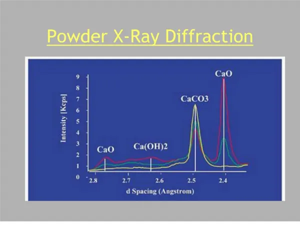
Uses: fingerprinting • Single or multi-phase Two different crystalline phases are present in this pattern – one in a very small amount NOT like spectroscopy. Whole patterns match.
Databases • To match, we need a very large database of powder patterns • ICDD (International Centre for Diffraction Data) Powder Diffraction File contains (2007) 199,574 entries (172,360 inorganic & 30,728 organic) • In ye olden days it was called JCPDS…(Joint Committee for Powder Diffraction Standards) and before that ASTM
ICDD Example Why d and not 2 ??
ICDD Good….
ICDD Bad….
Search/Match Search programs assist in identifying phase mixtures:
Inorganic Crystal Structure Database ICSD: ICSD
Fingerprinting.. Advantages: • relatively quick and easy, can be non-destructive • Problems: • need reliable standards - new phases will not be in the PDF • some things in the database are rubbish! • often need other (chemical) information to narrow down searches • not very sensitive - can “hide” up to 10% impurities (depending on relative “weights” – see later) • problems from preferred orientation, etc. • not much good for organics, organometallics.
Preferred Orientation Remember: we rely on a random orientation of crystallites. • When crystals are platey or needle-shaped (acicular) they will pack in a non-random fashion, preferentially exposing some planes to the incident radiation. Thus some diffraction peaks will be enhanced relative to others. This can also happen if a sample is packed down, or a thin film, etc. Brushite plates, SEM by Anna Fotheringham
Preferred Orientation Intensity mismatch – due to using single crystal So e.g. all (n00) peaks may be enhanced…
NaCl KCl Uses: different structures Even if two structures are the same (and they are chemically similar) differences can be observed: Peak positions (unit cell changes) and relative intensities (atoms) There is another major point here: K+ and Cl- are isoelectronic
Uses: different structures BUT, sometimes you can’t really see any changes on visual inspection… Zeolite A This often happens in “open” structures where there is space for change of light atoms
Uses: polymorphs Different polymorphs will have different powder patterns e.g. Zn S
Uses: polymorphs K3SO4F: tetragonal & cubic forms
Peak Broadening In an X-ray diffraction pattern, peak width depends on • the instrument • radiation not pure monochromatic • Heisenberg uncertainty principle • focussing geometry • the sample… - a crystalline substance gives rise to sharp lines, whereas a truly amorphous material gives a broad “hump”. What happens between the two?
Peak Broadening If crystal size < 0.2 m, then peak broadening occurs At <50nm, becomes significant. Why? Bragg’s law gives the condition for constructive interference. At slightly higher than the Bragg angle, each plane gives a “lag” in the diffracted beam. For many planes, these end up cancelling out and thus the net diffraction is zero. In small crystals, there are relatively fewer planes, so there is a “remanent” diffraction
Peak Broadening We can calculate the average size of the crystals from the broadening: Scherrer formula t is the thickness of the crystal, the wavelength, B the Bragg angle. B is the line broadening, by reference to a standard, so that where BS is the halfwidth of the standard material in radians. (A normal halfwidth is around 0.1o)
Peak Broadening Halfwidth: “Full width at half-maximum” - FWHM This can be different in different directions (anisotropic), so by noting which peaks are broadened we can also infer the shape of the crystals.
1050oC 30oC Uses: particle size determination Here we see particle size increasing with temperature
Particle size determination: Example Peak at 28.2° 2 with FWHM of 0.36° 2 Standard material has FWHM of 0.16° 2 = CuK = 1.540 Å 0.36 ° = 0.36 x /180 = 0.0063 rad 0.16 °= 0.16 x /180 = 0.0028 rad B = 0.0056 rad t = 255 Å = 0.0255 m
Particle size determinaton • An estimate, rather than an absolute value - also will be dominated by smallest particles. • Good for indication of trends. • A useful complement to other measurements such as surface area, electron microscopy etc.
Amorphous / micro-crystalline? It can be difficult to distinguish between an amorphous material and a crystalline sample with very small particle size. BUT the idea of such a small size “crystal” being crystalline doesn’t make sense! 5nm = 50Å = e.g. 10 unit cells Is this sufficient for long range order??
Unit cell refinement As the peak positions reflect the unit cell dimensions, it is an “easy” task to refine the unit cell. • 2d sin = and e.g. Thus if we can assign hkl values to each peak, we can gain accurate values for the unit cell We minimise the difference, e.g. This is known as “least squares” refinement. We will come back to this later.
Variable temperature/pressure Need special apparatus Here (see previous) we could follow a phase transition as we heated the sample up – following the change in unit cell parameters. J. M .S. Skakle, J. G. Fletcher, A. R. West, Dalton 1996 2497
BaTiO3 T/P Variable pressure hard to do: neutron diffraction (later) Much of these data actually from dielectric measurements. T. Ishidate, PRL (1997) 78 2397 S. A. Hayward, S. A. T. Redfern, H. J. Stone, M. G. Tucker, K. R. Whittle, W. G. Marshall, Z. Krist. (2005) 220 735.
Uses: more advanced Structure refinement – the Rietveld method A refinement technique, not determination Whole-pattern fitting - not just the Bragg reflections Needs a MODEL - pattern calculated from model, compared point-by-point with observed pattern. Originally developed (1967,1969) for use with neutron data - good reproducible peak shapes 1977 - first report of application to X-ray data Hugo Rietveld, b1932 http://home.wxs.nl/~rietv025/
Uses: Rietveld Refinement Here there was a similarity between the powder pattern of this phase and an existing one – also chemical composition similar. J. M. S. Skakle, C. L. Dickson, F. P. Glasser, Powder Diffraction (2000) 15, 234-238
Uses: more advanced • Quantitative phase analysis (how much of each) Naïve approach - relative intensity of peak maxima? - Consider mixture of Ba,Si,O - Ba component would scatter more than Si component (e.g. Ba2SiO4 c.f. SiO2) Thus uses Rietveld method and takes into account relative scattering from each crystalline phase
Summary Many different uses for powder X-ray diffraction! • Fingerprinting: identifying phases, distinguishing similar materials, identifying polymorphs, (following chemical reactions) • Indication of particle size from peak broadening • Unit cell refinement • Variable temperature/pressure measurements • Crystal structure refinement • Quantitative analysis