Download
1 / 41
410 likes | 560 Views
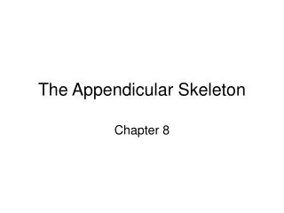
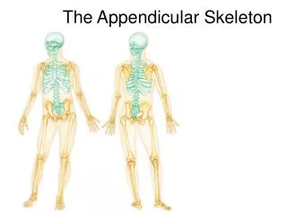
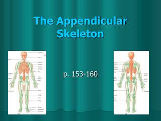
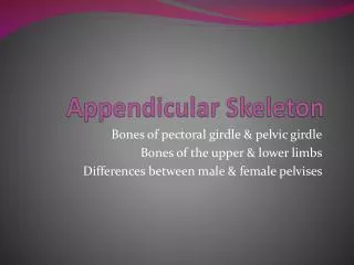
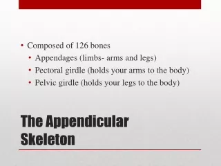
The Appendicular Skeleton. Honors A&P. The Clavicle. The Pectoral Girdle. ID your view. Anterior P osterior. ID the Acromion. 1 2 3 4 5 6 7 8 9. ID the Infraspinous Fossa. 1 2 3 4 5 6 7 8 9. ID the acromial end of the clavicle. 1 2 3 4 5 6 7 8 9.

E N D
The Appendicular Skeleton Honors A&P
ID your view • Anterior • Posterior
ID the Acromion • 1 • 2 • 3 • 4 • 5 • 6 • 7 • 8 • 9
ID the InfraspinousFossa • 1 • 2 • 3 • 4 • 5 • 6 • 7 • 8 • 9
ID the acromial end of the clavicle • 1 • 2 • 3 • 4 • 5 • 6 • 7 • 8 • 9
ID the psiform • 10 • 2 • 3 • 4 • 5 • 6 • 7 • 8 • 9
ID the trapezoid • 10 • 2 • 3 • 4 • 5 • 6 • 7 • 8 • 9
ID the deltoid tuberosity • 1 • 2 • 3 • 4 • 5 • 6 • 7 • 8 • 9
ID the greater tubercle • 1 • 2 • 3 • 4 • 5 • 6 • 7 • 8 • 9
ID the trochlea • 1 • 2 • 3 • 4 • 5 • 6 • 7 • 8 • 9
ID the radial tuberosity • 1 • 2 • 3 • 4 • 5 • 6 • 7 • 8 • 9
ID the ulnarstyloid process • 11 • 12 • 3 • 4 • 5 • 6 • 7 • 8 • 9
Is this a male or female pelvis? • Male • Female • Cannot be determined
ID the acetabulum. • 1 • 2 • 3 • 4 • 5 • 6 • 7 • 8 • 9
ID the iliac crest. • 1 • 2 • 3 • 4 • 5 • 6 • 7 • 8 • 9
ID the ischial spine • 1 • 2 • 3 • 4 • 5 • 6 • 7 • 8 • 9
Id the cuboid tarsal. • A • B • C • D • E • F • G
Id the navicular tarsal. • A • B • C • D • E • F • G
ID the lateral malleolus • 1 • 2 • 3 • 4
Articulations (Joints) • Articulations – wherever 2 bones meet • Classified by structure • Fibrous (binding connective tissue) • Cartilaginous (binding connective tissue) • Synovial (permit free movement) • Classified by function • Synarthrosis (Immovable) • Amphiarthrosis (slightly movable) • Diarthrosis (synovial joints)
Synathrosis (no movement) • Sutures • Bones of the skull • Gomphosis • Ligament bonds tooth w/in bony socket • Cartilaginous • Connection between 1st rib and sternum
Amphiarthroses (Slightly Movable) • Syndesmosis • Fibrous joint connected by ligament • Ex. Distal articulation between tibia and fibula • Symphysis • Bones joined by disk of fibrocartilage • Ex. Vertebrae, between pubic bones
Diarthrosis (Synovial Movement) • Bound by joint capsule and contains synovial fluid • Types of movement: • Gliding – 2 opposing surfaces slide past one another (carpals, tarsals) • Angular • Rotation • Special Movement
Angular Movements • Angular Motion • Flexion – reduces angle between articulating elements • Extension - increases angle between articulating elements • Adduction – moving towards midline • Abduction – moving away from midline • Circumduction – loop motion
Rotational Movements • Rotational
Special Movements • Inversion- turns sole of foot inward (opp-eversion) • Dorsiflexion- ankle flexion (plantar flexion pointed toe) • Opposition – grasping (thumb/fingers toward hand) • Protraction - move anterior across horizontal plane (opp retraction) • Elevation – move superior (opp depression)
Structural Classification of Synovial Joints • Gliding – flat surfaces slide past one another • Ends of clavicles • Between carpals & tarsals • Between vertebrae • Hinge – angular movement in a single direction • Occipital bone and atlas • Elbow, knee, ankle • Interphalangeal joints • Pivot – permit rotation only • Atlas and axis • Proximal radius and ulna • Ellipsodial – angular motion occurs in 2 planes • Radius w/proximal carpals • Phalanges w/metacarpals (and metatarsals) • Saddle- permits angular motion but prevents rotation • thumb • Ball and socket - round head rests within depression • Shoulder • hips