Download
1 / 34
380 likes | 714 Views
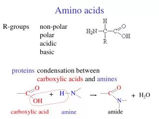
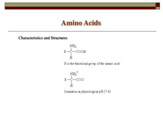
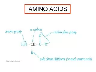
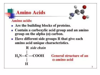
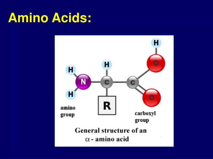
Amino Acids:. Peptide Bond. * Elimination of water upon formation. * Peptide bond is flat. Non-polar. Aromatic. Residues (R’s):. + Charged. Polar, uncharged. - Charged. Groups of Amino acids:. Amino Acid Types:. * Water-loving ( polar ): - Charged: Has an NH3+, or COO-

E N D
* Elimination of water upon formation. * Peptide bond is flat.
Non-polar Aromatic Residues (R’s): + Charged Polar, uncharged - Charged Groups of Amino acids:
Amino Acid Types: • * Water-loving (polar): • - Charged: Has an NH3+, or COO- • - not charged: Typically, contain an oxygen. • * Water-’hating’ (non-polar): • Typically, R’s do not contain any oxygen, or Aromatic. • * Small – Flexible (Glycine)
H H O Amino acid: H N C C O H
Let’s build a peptide (short protein): Step One: Prepare amino acid A.Receive a page of an amino acid. Draw in the atoms of the “R” group. B.Color the R group by category: Charged positively –Blue Charged negatively –Red Polar uncharged – Black Non-polar, Aromatic –Yellow
Step Two: Connecting by peptide bond 3. Find the –OH group for the COOH and the –H from the NH2 side. Circle them. 4.With your neighbor: One cutsout the OH, and the other the –H. 5. Bring the C=O and N-Htogether with a bond. Use Clip to connect.
We just made a class – size peptide. It has two edges: COOH and a NH2. The 3-dimentional structure will be determined by the categories of the amino acids (coloring)..
How many 3-digit numbers are possible using 2,3,4? Explain. • Like-wise, how many 3-long proteins can be made at random from just 3 amino acids: Alanine, Histidine, Serine? • Repeat but using all possible 20 known amino acids.
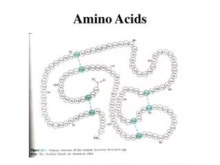
Diversity of Structures creates diversity of functions. .. In fact, proteins range between 20 to 1000 amino acids (average ~ 100 amino acids). Which means ______ random combinations!
Diversity of Amino Acid Sequences generated diversity at least as large of 3D structures. Lamin
..but how does the amino acid sequences (primary structure) determine the 3D structure? The side chains interact:
Types of interactions: Disulfide Bond Hydrogen Bond Electrostatic VanDerWaals (hydrophobic)
Hydrogen Bond • Occurs between a hydrogen ‘donor’ and ‘acceptor’: • Donor = Has a partially charged H. • Acceptor = Has a partially negatively charged.
Electrostatic Interactions: • Opposite charge – attraction • Same charge = repultion
Disulfide Bonds An actual covalent bond. Is relatively strong, but not very common.
Van der Waals Interactions * Between atoms that are close enough: Attraction between electrons of one atom to the nucleus of another. * The weakest of all, but numerous. * Associated with hydrophobic exclusion.
Hydrophobic Exclusion: Rejection by the surrounding water forces R groups to come together, minimizing the contact with water. A MAJOR force in protein folding into domains. Hydrophobic folding
Protein folding - simplified Collagen PPi hydrolase Antibody Insulin
Splitting Water 1 out of every 550 million water molecules can spontaneously break into and OH- (‘Hydroxyl’): H2O H+ (‘proton’) + OH- (‘Hydroxide’) H+: No electrons, only 1 proton) OH_: One Extra e-.
Acids: Chemicals that dissociate in water to H+ ions. Bases: Chemicals that dissociate in water and absorb H+ and therefore reduce the acidity H+ Concentration is measured in molarity: Mole = 6.02x10²³. 1 Molar = one mole per liter. Pure water contain 10-7moles/liter.
pH: Power of Hydrogen * pH is the (– logarithm) of H+ concentration. bases are low in H+ (high in OH-). * Accordingly pure water is: -log (10-7) = 7 * Therefore, a change in one unit of pH in fact means a ‘jump’ of ten fold the concentration. 14
pH: Power of Hydrogen pH ranges from 1 to 14: 1 2 3 4 5 6 7 8 9 10 11 12 13 14 High H+ Low H+ Neutral Acids Bases pH of solutions is typically examined with indicator dies Add examples of the household materials to the scale
The pH Scale Measures the Concentration of H+ ions. Acidic Basic (Colors of an indicator) Low pH High pH
Demo: Adding acid to a protein solution from Egg white. a. Describe: What happened? b. What other conditions would give the same effect? So, acids can be harmful to proteins
Many of the forces that hold a protein fold together are hydrogen bonds: O O-H···· H+ Hydrogen Bonds are interrupted by acids and sometimes by bases.
To maintain the pH of a body solution we use substances that can stabilize the pH. They are called buffers. The most important buffer in our body (pH 7.4) is carbonate: H2O + CO2 H2CO3 HCO3-+ H+ So, is this an acid or a base?
Exercise: Use Toobers and Tacks to demonstrate how the primary sequence of a protein can determine the 3-dimentional structure of a protein.
Primary Structure (AGVTDPG) Secondary Tertiary (3D)