Download
1 / 1
10 likes | 169 Views
Distribution of nerve fibres in bovine and human mucosal associated lymphoid tissues. V.Defaweux 1 , G.Dorban 1 , N.Antoine 2 , J.Piret 2 , A.Gabriel 3 , O.Jacqmot 3 , S.Flandroy 1 , D.Zorzi 1 , E.Heinen 1

E N D
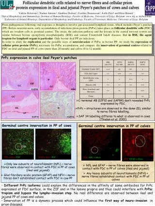
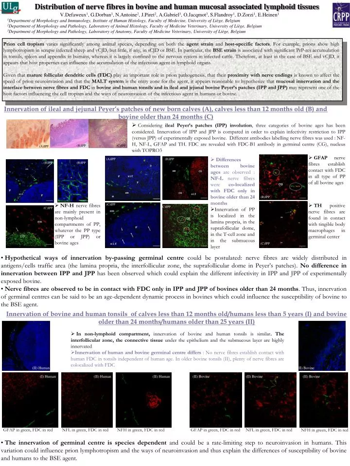
Distribution of nerve fibres in bovine and human mucosal associated lymphoid tissues V.Defaweux1, G.Dorban1, N.Antoine2, J.Piret2, A.Gabriel3, O.Jacqmot3, S.Flandroy1, D.Zorzi1, E.Heinen1 1Department of Morphology and Immunology, Institute of Human Histology, Faculty of Medecine, University of Liège, Belgium 2Department of Morphology and Pathology, Laboratory of Animal Histology, Faculty of Medicine Veterinary, University of Liège, Belgium 3Department of Morphology and Pathology, Laboratory of Anatomy, Faculty of Medicine Veterinary, University of Liège, Belgium Prion cell tropism varies significantly among animal species, depending on both the agent strain and host-specific factors. For example, prions show high lymphotropism in scrapie infected sheep and vCJD, but little, if any, in sCJD or BSE. In particular, the BSE strain is associated with significant PrP-res accumulation in tonsils, spleen and appendix in humans, whereas it is largely confined to the nervous system in infected cattle. Therefore, at least in the case of BSE and vCJD, it appears that host properties can influence the accumulation of the infectious agent in lymphoid organs. Given that mature follicular dendritic cells (FDC) play an important role in prion pathogenesis, that their proximity with nerve endings is known to affect the speed of prion neuroinvasion and that the MALT system is the entry zone for the agent, it appears reasonable to hypothesize that mucosal innervation and the interface between nerve fibres and FDC in bovine and human tonsils and in ileal and jejunal bovine Peyer’s patches (IPP and JPP)may represent one of the host factors influencing the cell tropism and the ways of neuroinvasion of the infectious agent in humans or bovine. Innervation of ileal and jejunal Peyer’s patches of new born calves (A), calves less than 12 months old (B) and bovine older than 24 months (C) • Considering ileal Peyer’s patches (IPP) involution, three categories of bovine ages has been considered. Innervation of IPP and JPP is compared in order to explain infectivity restriction to IPP (versus JPP) of experimentally exposed bovine. Different antibodies labelling nerve fibres was used : NF-H, NF-L, GFAP and TH. FDC are revealed with FDC-B1 antibody in germinal centre (CG), nucleus with TOPRO3 (B)IPP (C)IPP (A)IPP • GFAP nerve fibres establish contact with FDC in all type of PP of all bovine ages • Differences between bovine ages are observed : NF-L nerve fibres were co-localized with FDC only in bovine older than 24 months • Innervation of PP is localized in the lamina propria, in the suprafollicular dome, in the T-cell zone and in the submucous layer (A)IPP (B)IPP (B)IPP (B)IPP (B)JPP • NF-H nerve fibres are mainly present in non-lymphoid compartments of PP, whatever the PP type (IPP or JPP) or bovine ages (C)IPP • TH positive nerve fibres are found in contact with tingible body macrophages in germinal center (C)IPP (C)IPP (C)JPP • Hypothetical ways of innervation by-passing germinal centre could be postulated: nerve fibres are widely distributed in antigens/cells traffic area (the lamina propria, the interfollicular zone, the suprafollicular dome in Peyer’s patches). No difference in innervation between IPP and JPP has been observed which could explain the different infectivity in IPP and JPP of experimentally exposed bovine. • Nerve fibres are observed to be in contact with FDConly in IPP and JPP of bovines older than 24 months. Thus, innervation of germinal centres can be said to be an age-dependent dynamic process in bovines which could influence the susceptibility of bovine to the BSE agent. Innervation of bovine and human tonsils of calves less than 12 months old/humans less than 5 years (I) and bovine older than 24 months/humans older than 25 years (II) • In non-lymphoid compartment, innervation of bovine and human tonsils is similar. The interfollicular zone, the connective tissue under the epithelium and the submucous layer are highly innervated • Innervation of human and bovine germinal centre differs : No nerve fibres establish contact with human FDC in tonsils independent of human age. In older bovine tonsils (II), plenty of nerve fibres are colocalized with FDC (II) Human (II) Bovine (I) Human (II) Human (II) Human (II) Bovine (II) Bovine (II) Bovine GFAP in green, FDC in red NFL in green, FDC in red NFH in green, FDC in red GFAP in green, FDC in red NFL in green, FDC in red NFH in green, FDC in red • The innervation of germinal centre is species dependent and could be a rate-limiting step to neuroinvasion in humans. This variation could influence prion lymphotropism and the ways of neuroinvasion and thus explain the differences of susceptibility of bovine and humans to the BSE agent.