Download
1 / 18
180 likes | 339 Views
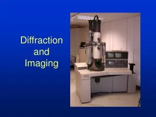
Diffraction Enhanced Imaging at the UK Synchrotron Radiation Source. M.Ibison, K.C.Cheung, K.Siu, C.J.Hall, R.A.Lewis, A. Hufton, S.J.Wilkinson, K.D.Rogers, A.Round. Principal Objectives of DEI Development Activities at the SRS.

E N D
Diffraction Enhanced Imaging at the UK Synchrotron Radiation Source M.Ibison, K.C.Cheung, K.Siu, C.J.Hall, R.A.Lewis, A. Hufton, S.J.Wilkinson, K.D.Rogers, A.Round
Principal Objectives of DEI Development Activities at the SRS • reduce the proportion of available time spent on alignment, in preparation for useful imaging • increase reliability and stability in mechanics and software, to improve image quality • approach a ‘turn-key’ facility for DEI users without need for expertise in the detailed method. • Also:- • obtain useful experience for designing 2nd generation DEI on higher-energy SRS station.
DEI System – Station 7.6 • High precision optics required • 2-crystal monochromator and 2-crystalanalyser • Si 311 crystals give sharper x-ray extinction • Higher contrast, higherresolution images.
X-Ray Optics AlignmentLaser • red beam (l = 623nm), <1 mW output • compact (2.5cm x 1cm), power source = 2 x 1.5V batteries • micrometer adjustments, Vertical & Horizontal angle & displacement • spot of 2mm x 1mm at working range of 2m
Monochromator and 4-Crystal Alignment using Laser Assistance Monochromator Alignment Analyser Alignment NOTE: Vertical Spacing between crystals exaggerated for clarity.
Laser Method for Determination of Motor Drive Calibration Factors
Laser Method for Determination of Motor Drive Calibration Factors
Ionisation Chamber for X-Ray Beam Location • polymer window (5cm x 1cm) • thickness = 70mm • atmospheric pressure
Silicon p.i.n. PhotoDiodefor DEI Alignment • Sensitive Area: 3.5mm x 3.5mm • Thickness (effective): 250mm • Window: 10mm Al foil (for 14keV X-rays)
Medical Applications of DEI: Mouse Feet Study Normal Diseased Normal Diseased Refraction Images Absorption Images • vertical view • through sole of foot
Mouse Feet Study (2) Refraction Images Normal Diseased Normal Diseased • horizontal view • through side of foot Absorption Images
DEI Insect Studies Beetle Earwig Refraction Image Absorption Image
Examples of CT Reconstruction Input to Reconstruction = Set of Projections (Sinogram) Results of Reconstruction = Cross-Section (Slice)
Effect of Filtering on Reconstructed Image Unfiltered Filtered
Volume Visualisation • 3-D Rendering of a Mouse Liver, based on CT dataset (ELETTRA) • Uses Volume/Surface modelling features of software packages
Second-Generation DEI System:Some Design Considerations • higher energy and greater flux (on wiggler Station) - better penetration, lower subject dose • channel-cut crystals - facilitate alignment, reduce drift • rigidity and anti-vibration built into support structure • mountings optimize use of existing framework - station sharing remains feasible • vacuum enclosure of the monochromator - avoids convection currents and ozone damage risk • cooling provision for the 1st crystal - highest heat loading from ‘white’ beam.
Acknowledgements The authors would like to thank:- Medical Research Council - for funding this research programme Giuseppe Salvini and Janet Groves (CLRC Daresbury) - design and construction of the p.i.n. diode device Greg Johnson (CLRC Rutherford Appleton Laboratory) - design and implementation of DSP reconstruction system Andrew Mather (Liverpool University) - Java implementation of the FBP reconstruction software.