Download
1 / 9
90 likes | 104 Views
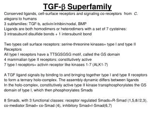
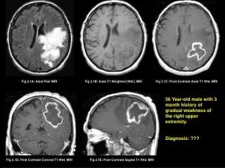
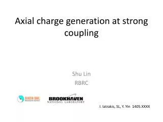
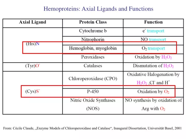
Hemoproteins: Axial Ligands and Functions. From: Cécile Claude, „Enzyme Models of Chloroperoxidase and Catalase“, Inaugural Dissertation, Universität Basel, 2001. F8390. Metalloproteins: Structure and Function Introduction 1.1. Metalloproteins: Functions in Biological Chemistry

E N D
Hemoproteins: Axial Ligands and Functions From: Cécile Claude, „Enzyme Models of Chloroperoxidase and Catalase“, Inaugural Dissertation, Universität Basel, 2001
F8390 • Metalloproteins: Structure and Function • Introduction • 1.1. Metalloproteins: Functions in Biological Chemistry • 1.2. Some fundamental metal sites in metalloproteins • 2. Mononuclear zinc enzymes: Carbonic anhydrase • 3. Metalloproteins reacting with oxygen • 3.1. Why do aerobic organisms need metalloproteins? • 3.2. Oxygen transport proteins & Oxygenases • 3.2.1. Hemoglobin, Myoglobin Cytochrome P450 • 3.2.2. Hemerythrin & Ribonucleotide Reductase R2 & • Methane monooxygenase diiron subunits • 3.2.3. Hemocyanin & Tyrosinase • 4. Electron transfer proteins • 4.1. Iron-sulfur proteins • 4.2. Blue copper proteins • 5. Conclusion
Modification of the FeII/FeIII redox potential by the protein environment Strong oxidants FeII (Red.) stable FeIII (Ox.) stable Strong reductants Hemoprotein proximal ligand Em for FeII/FeIII (mV) FeIII/FeII (aq.) FeIII/FeII - +770 Human hemoglobin FeIII/FeIIHis +150 Microperoxidase11-CO FeIII/FeIIHis +100 Chloroperoxidase FeIII/FeIICys- -150 NO synthase neuronal FeIII/FeIICys- -250 Horse-radish peroxidase FeIII/FeIIHis -280 Cytochrome P450 2C5 FeIII/FeIICys- -330 Catalase FeIII/FeIITyr- -460 Source: C. Capeillere-Blandin, D. Matthieu & D. Mansuy, Biochem. J. 2005, 392, 583-587 Different metalloproteins need different redox potential for their function. Cytochrome P450 needs to access the unusual oxidation state Fe(V) to be able to oxidize even unreactive substrates. Therefore, it uses the negatively charged cysteine ligand which donates electrons to Fe and stabilizes the high oxidation state.One of strategies that proteins employ to modify the redox potentialis using different proximal ligands.
Examples of Cytochrome P450 substrates • Hydroxylation at: • aliphatic carbons • -aromatic carbons • double bonds steroid hormone local anesthetic -heteroatoms carcinogen from fungi antibiotic Alkaloid from Taxus brevifolia, potent anti-cancer drug
Cytochrome P450cam (Campher-5-monooxygenase; pdb-code 1T86) access for substrate and O2
Catalytic cycle of cytochrome P450cam Substrate RH binds into the hydrophobic pocket and pushes H2O out from coordination site E ~ -0.17 V Low-spin FeIII Em≤ -0.3 V e- from putidaredoxin Redox parnter of P450cam: putidaredoxin (Fe-S protein) Em≈ -0.2 V O2 binds to the empty coordination site e- from putidaredoxin http://www.cup.uni-muenchen.de/ac/kluefers/homepage/L_bac.html
Conclusion • In many cases, metalloproteins use the same or similar active site • for different purposes. • The strategies to confer a particular activity to a given site include • Allowing/disallowing access of substrates to the active site • (including the dynamics of diffusion of substrate/product) • Modifying the electrostatic potential by mutating the amino acids • coordinated to the metal or surrounding the binding pocket
Practical training - Download from the pdb database the structures of bacterial cytochrome P450cam 1t86 and 1dz8 http://www.rcsb.org/pdb/home/home.do • - Display the structures using VMD • - Use the command „chain A“ in Graphics/Representation“ to display only the monomer A • - Use the command „chain A and resname HEM“ in Graphics/Representation“ to highlight the heme group • - Observe whether the two crystal structures contain the campher and/or oxygen molecule trapped near the active site • - Use the command „chain A and resname CAM“ in Graphics/Representation“ to highlight the campher molecule • Use the command „chain A and resname OXY“ in Graphics/Representation“ to highlight the O2 molecule • Examine how the two carboxylate groups of heme are anchored in the protein backbone • Examine how the campher substrate is fixed in the acess channel