Download

1 / 25
280 likes | 461 Views
THE OXFORD CLASSIFICATION OF IgA NEPHROPATHY: SINGLE CENTRE EXPERIENCE. Petrusevska G., Jasar G., Grcevska L., Kostadinova S., Bogdanovska M.*, Nikolov V., Polenakovic M. Macedonia. INTRODUCTION.

E N D
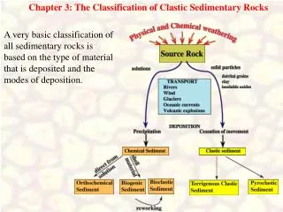
THE OXFORD CLASSIFICATION OF IgA NEPHROPATHY: SINGLE CENTRE EXPERIENCE Petrusevska G., Jasar G., Grcevska L., Kostadinova S., Bogdanovska M.*, Nikolov V., Polenakovic M. Macedonia
INTRODUCTION • The diagnosis of IgA nephropathy, the commonest glomerular disease worldwide, is defined by the presence of mesangial IgA-dominant immune deposits within glomeruli in the absence of systemic disorders1,2,3,4,6. • This criterion is the reason for a wide range of histological changes in IgA nephropathy (IgAN) and a different outcome of the renal disease 1,5,7.
INTRODUCTION • Biopsy appearances may range from: • normal glomeruli by optical microscopy; • severe crescentic glomerulonephritis; • sclerosing glomerulonephritis. • Some biopsies present dominant diffuse mesangial proliferation and the other focal proliferative changes with different degrees of tubulointerstitial changes.
INTRODUCTION • Thus many authors in the past tried to make a usefull classification, comparing the histopathological findings with clinical features and the outcome of the disease7,8,9,10. • None has achieved widespread acceptance, because of a lack of definitions, and inclusion of both active and chronic lesions in the definitions of single categories. • There is continuing discussion as to wheather pathological features seen on renal biopsy contribute additional prognostic information in addition to the clinical features.
INTRODUCTION • The aim of the new Oxford classification 11,12,13 was to identify specific pathological features that can predict the risk of progression of renal disease in IgAN. • Six of the pathological variables were identified as having an independent value in predicting renal outcome: 1) the mesangial hypercellularity score; percentage of glomeruli showing, 2) segmental sclerosis, 3) endocapillary hypercelullarity or 4) cellular/fibrocellular crescents;5) percentage of tubular atrophy/interstitial fibrosis and 6) arteriosclerosis score. • These features were recommended to be taken into account for predicting an outcome independent of the clinical features both at the time of presentation and during follow-up.
AIMS • The aim of our study was: • To define the presence of the histopathological variables recommended by Oxford classification in Macedonian patients with diagnosed IgAN. • To corelate the value of these pathologic variables and outcome of the disease in this group of patients with IgAN. • To determine the survival in two groups of patients: those with nephrotic syndrome (defined as proteinuria> 3g/daily) and immunosupression and the other group of patients, with mild clinical features and without immunosupression.
PATIENTS AND METHODS • The study had a retrospective character on cases with biopsy-proven IgAN at the Institute of pathology and Laboratory for cytology and histopathology, Institute of oncology, Medical faculty in Skopje, admitted at the University Nephrology Department. • Histological variables were analysed according to the histopathological reports because of long period survey analysis. • The diagnosis of IgAN includded: 1) demonstration of mesangial IgA deposition by DIF in isolation or as the predominant immunoglobulin and the lack of clinical or serological evidence of systemic lupus erythematosus, Henoch-Scoenlein purpura, or liver disease; 2) standard histological analysis of the renal biopsies on HE, PAS, Trichrome Mason, Silvermethenamine-Jones stained sections; and 3) Electron microscopy performed in 6% of the patients, and semithin sections in about 70%.
SELECTION OF THE PATIENTS • Histopathological reports and the clinical data at the onset and during follow-up were obtained from the in-patient and out-patient files of the patients at the University Nephrology Department which covers renal disease patients in all area of R. Macedonia (about 2,000,000 people). • Ninety eight adult patients were classified as having IgA nephropathy during the period 1976-2006. • 40 adult patients (25 male, 15 female,>15 years) were selected for the study, patients with more than 10 glomeruli at optical microscopy and with a follow-up of more than 3 years. • We used four pathological variables proposed by the Oxford Classification as it is presented in Table 1.
Table 1.: Definition of pathological variables used in the classification of IgAN.
Statistics • Statistics included: • Student`s T test, Spearman`s test and Mann-Whitney U Wilcoxon test to compare histological scoring and duration of follow-up without end-stage renal disease. • Histopathological score and survival were compared separately 1) in all 40 patients, independently of clinical signs and treatment; 2) 12 patients with nephrotic syndrome (defined as proteinuria> 3g/daily) and immunosupression were analysed separately, and 3) the other 28 patients, with mild clinical features and without immuniosupression, were also analysed separately. • The Kaplan-Mayer test was performed to determine the survival of the patients.
Table 2: RESULTS: Histological findings in whole group of 40 pts
RESULTS: Overall survival of the whole group of 40 pts) • The average survival of the whole group was 10.8+/-7.47 years (M+/-SD). • Twenty from forty (50%) of the patients experienced end stage renal disease after a period of 3-12 years. • Pathological variables and follow-up of all 40 patients according to the recommendation of the Oxford classification are presented at Table 3. • Mesangial hypercelullarity was confirmed to be associated with the renal outcome (p=0.047), as well as glomerular sclerosis (p=0.009), endocapillary hypercellularity (p=0.001) and tubular atrophy/interstitial fibrosis (p=0.045)
Table 3: Influence of the histological variables to the renal outcome
Results of Group I patients (without nephrotic syndrome) • This group consisted of 28 patients with clinically normal renal function, without hypertension and nephrotic syndrome at presentation. Thirteen from 28 (46.1%) of these patients, in contrast to mild clinical features at presentation, presented end-stage renal disease during follow up. • Mesanagial hypercelullarity didn`t correlate with renal survival in this separated group of patients (p=0.053 Mann Whitney, p=0.49 Spearman test). Segmental glomerulosclerosis significantly correlated with the outcome of the disease (p=0.007 Mann Whitney, p=0.006 Spearman test). Endocapillary hypercelullariry (p=0.351, p=0.32) as well as tubular atrophy/interstitial fibrosis (p=0.071, p=0.105) did not correlated with the renal outcome.
Table 3: Influence of the histological variables to the renal outcome
Results of Group II patients (with nephrotic syndrome) • Group II consisted of 12 patients with severe clinical features at presentation and necessary immunosuppression. • Seven from 12 (58.3%) developed terminal phase of chronic renal failure during follow-up. • Renal survival was not associated with mesangial hypercelullarity (Man Whitney p=0.606, Spearman test p=0.607); with glomerular sclerosis (p=0.343, p=0.302) and endocapillary hypercelullarity (p=0.53; p=0.533), • But it was associated with the degree of chronic tubulointerstitial changes (p=0.018, p-0.016).
Table 3: Influence of the histological variables to the renal outcome
Survival of both groups of patients with IgA nephropathy • It can be seen that there was noted significant difference in survival between those two group of patients. • Glomerular sclerosis was a poor prognostic pathologic variable in the patients without nephrotic syndrome, and tubulointerstitial changes in ones with nephrotic syndrome.
DISCUSSION • Many authors have tried to correlate clinical signs and histological features in IgAN7,8,9,10, 14,15,16. • Gross hematuria was frequently associated with mild glomerular lesions and a favourable clinical course; on the other hand, the presence of heavy proteinuria at the onset was frequently associated with severe histology and progressive renal disease.
DISCUSSION • Severe histology was also associated with already decreased renal function and/or hypertension at the time of biopsy. Many authors believe that the histologic type of glomerular lesions appears to be the best predictive index in IgAN. • The folowing factors were regarded as histological parameters of progressive damage in IgAN: severe mesangial proliferation, frequent sclerotic glomeruli, crescents higher proportions of glomerular adhesions, vascular sclerosis and marked interstitial fibrosis (8,9,14). They are all causally correlated with each other in portending a poor prognosis.
DISCUSSION • Several histologic grading systems have been used in the past8,17,18. These pathological systems used to classify renal lesions in IgAN, can be divided into two groups: lumped and split. • The lumped systems assess the overall severity of histological lesions, found in glomerular, tubular, interstitial and arteriolar compartments, as in the widely-used classification of Lee and Haas8,18. The split systems use semiquantitative severity grading of lesions in each of the four compartments and permit the elaboration of a global or aggreagate score for each compartment
DISCUSSION • Lesions of focal and segmental hyalinosis and sclerosis were described as very specific for progression and so in our previous report we measured the sclerotic glomerular area and combined it with the semiquantitaive score of the tubulointerstitial changes in order to predict the progression19. • In general, the agreement among various classifications is only reached when considering renal disease already progressed to sclerosis.
DISCUSSION • The new Oxford classification11,12,13 documented by univariate and multivariate analysis that the following lesions resulted as independently predictive of clinical outcome: mesangial hypercelullarity score, endocapillary hypercelullarity, segmental glomerulosclerosis and tubular atrophy/interstitial fibrosis. Necrotizing and crescentic lesions were not evaluated because of their rarity. Our results taking into consideration only those four histological variables presented the same results, a higher total score – a more progressive disease. • Our group consisted of 40 patients of different age, different clinical presentations and different treatment depending on the presence of nephrotic syndrome, and these four variables proposed by the Oxford classification were important for the outcome of the disease.
In memory to • Prof.dr. Georgi Zografski, pathologist, Laboratory for cytology and histopathology, Institute of Oncology Geographic position of R. Macedonia and the church of St. Kaneo in Ohrid