Download
1 / 37
380 likes | 574 Views
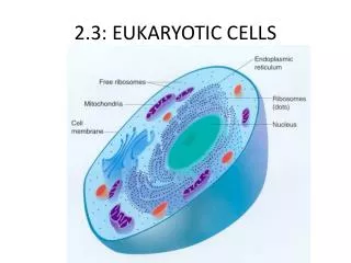
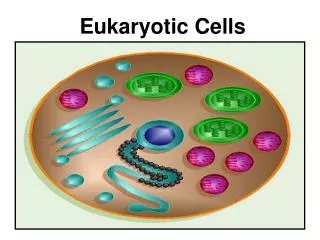
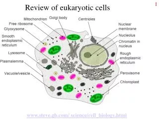
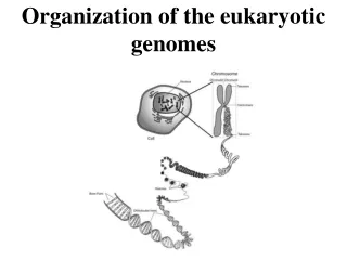
II. INTERNAL ORGANIZATION OF EUKARYOTIC CELLS. NUCLEOLUS. Cell nuclei and nucleoli in the liver. Light micrograph Haematein-eosin staining. The nucleolus in electron micrographs. Spherical structures within the nucleus, ~ 1-2 m m.

E N D
Cell nuclei and nucleoli in the liver. Light micrograph Haematein-eosin staining
The nucleolus in electron micrographs Spherical structures within the nucleus, ~1-2 mm. In interphase cells: nucleolus is normally not readily recognisable or absent, except in neurons, where a large, prominent nucleolus is present.
MAIN PARTS OF THE NUCLEOLUS: NO: nucleolar organizer region of those chromosomes possessing nucleolar genes PF: pars fibrosa, dense fibrillary component PG: pars granulosa, or nucleolonema: ribonucleo- protein particles NAC: nucleolus-associated chromatin
FUNCTION OF THE NUCLEOLUS: It produces the ribosomes. Main steps: - transcription of nucleolar genes code for ribosomal RNA occurs at the nucleolar organizer - ribosomal proteins are syntesized in the cytoplasm - RNA-protein assembly takes place in pars fibrosa - ribonucleoprotein particles accumulate in pars granulosa and undergo maturation process - completed ribosomal subunits are transported through the nuclear pores into the cytoplasm where they carry out their function
Nissl staining of neuronal cell bodies visualizes stacks of rER at light microscopic level
Endoplasmic reticulum (ER) in EM SER RER Nucleus Nuclear membran
Region of peptide synthesis mRNA Large subunit Small subunit Exit of new peptide Structure of a ribosome
Free and ER-bound ribosomes mRNA encoding a cytosolic protein pool of ribosomal subunits in cytosol mRNA encoding a protein targeted to ER ER membrane
The sarcoplasmic reticulum: the special form of SER in skeletal muscle Function: Ca ion traffic for the contraction
Functions The endoplasmic reticulum plays key role in the synthesis of several types of molecules. ER and the Golgi complex exert correlated, coordinated activity. Proteins are synthesized on RER-bound ribosomes (signal sequences!). After completion of the synthesis several types of proteins are submitted to post-translational modifications. Proteins are then further transported to the Golgi apparatus. Phospholids and fatty acids are synthesized and metabolised in the SER Further roles: Liver: glycogen synthesis, detoxication Adrenal gland: synthesis of steroid hormons Skeletal and heart muscle: storage of calcium ions.
GOLGI COMPLEX: RECONSTRUCTION FROM EM
FUNCTIONAL CONNECTION BETWEEN THE RER AND GOLGI MEMBRANES
LYSOSOMES: Acidic organelles that contain a battery of degradative enzymes. Membrane-limited organelles, degrade proteins, particles taken up by the cell, or the cell’s own degenerating organelles. Types: - primary lysosomes: spherical, do not contain particles or membrane debris, do contain acid hydrolases (phosphatases, nucleases, proteases, etc.) - secondary lysosomes: larger, irregularly shaped, do contain debris that is being digested. PEROXISOMES, GLYOXISOMES: Small membrane-limited organelles. Contain enzymes that degrade fatty acids and amino acids. A product of these reactions is hydrogen peroxide, a corrosive substance. Catalase of peroxisomes degrades the dangerous molecule.
Modification of proteins in the Golgi apparatus: - alteration of amino acid side chains - addition of saccharide residues - remodeling of oligosaccharides - specific proteolytic cleavages - formation of disulphide bonds - assembly of multiprotein complexes
Types of secretion : - continuous: proteins are sorted in the trans-Golgi reticulum into vesicles that immediately fuse with the cell membrane (example: collagen secretion of fibroblast cells in connective tissue) - regulated: the release of these proteins is initiated by different neural and hormonal stimuli. The exocytosis is triggered by a rise in the intracellular calcium level (example: hormone production of endocrine glands).
Mitochondrial types according to the form of the inner membrane Mitochondria with cristae: Cristae: thin folds which project into the interior of the mitochondrion. Occurence: most cell types.
Tubular mitochondria: the inner membrane projections are relatively broad tubes. Occurence: steroid hormone synthesizing cells (adrenal cortex, Leydig cells in testis)
Mitochondria with prisms: some of the cristae form triangular tublike structures (astrocytes) Sacculus-type mitochondria: the inner membrane tubuli are decorated with pearl-like broadenings (adrenal cortex)
Origin of mitochondria: symbiosis of prokaryotes Mitochondria arise exclusively by division, no de novo genesis! Mitochondria containDNA, RNA and ribosomes: they can divide by themselves. Nuclear DNA codes for 600-1000 mitochondrial proteins, while mtDNA contains the code for 8-13 ( ~ 1 %). Import of proteins synthesized in the cytosol : by a translocator-protein complex in the mitochondrial membrane. Mitochondria are always inherited from the mother (the mitochondria of the sperm are unable to get into the oocyte during fertilization - determination of female site pedigree).
Negative staining of the inner mitochondrial membrane: cristae contain elementary particles
STRUCTURE OF THE INNER MITOCHONDRIAL MEMBRANE 1. Enzymatic coupling factors 3. Enzymes of respiratory chain 4. Cytochrome C molecules 5. Outer chamber 6. Lipid bilayer 1. Outer membrane 2. Inner membrane 3. Crista 4. Head of elementary particle 5. Stalk of elementary particle
Functions Main function: „power plant” of the cell: synthesis of ATP by the enzymatic oxidation of amino acids, fatty acids and glucose. Functions of the intramitochondrial compartments: Outer membrane outer cover, protein import, pores Intermembrane space cytochromes, apoptosis factors Inner membrane Respiratory chain, ATP-synthesis, protein import, metabolite transport Matrix Citrate cycle, β-oxidation of fatty acids, mtDNA replication, proteine biosynthesis, haem synthesis, urea cycle