Download
1 / 19
700 likes | 2.29k Views
Labeled Immunoassays Part 4. Fluorescent & Chemiluminescent Immunoassays. Fluorescence Immunoassay. In 1944 it was demonstrated that antibodies could be labeled with molecules that fluoresce. These fluorescent compounds are called fluorophores or fluorochromes.

E N D
Labeled ImmunoassaysPart 4 Fluorescent & Chemiluminescent Immunoassays
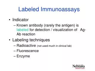
Fluorescence Immunoassay • In 1944 it was demonstrated that antibodies could be labeled with molecules that fluoresce. • These fluorescent compounds are called fluorophores or fluorochromes. • They have the ability to absorb energy from an incident light and convert that energy into light of a longer wavelength and lower energy as the excited electrons return to the ground state. • Each Fluorophore has a characteristic optimum absorption range. • The time interval between absorption of energy and emission of fluorescence is very short and can be measured in nanoseconds.
Characteristics of Fluorescent molecule Ideally, a fluorescent molecule should: • Exhibit high intensity, which can be distinguished easily from background fluorescence, • It should also be stable, • and have a high molar extinction coefficient (a measurement of how strongly a chemical species absorbs light at a given wavelength)
Types of Fluorescent molecules • The two compounds most often used are: • Fluorescein • and Rhodamine, • because these can be readily coupled with antigen or antibody.
Types of Fluorescent molecules • Fluorescein • absorbs maximally at 490 to 495 nm • and emits a green color at 517 nm. • It has a high intensity, • good photostability, • and a high quantum yield. • Tetramethylrhodamine • absorbs at 550 nm and • emits red light at 580 to 585 nm. • Because their absorbance and emission patterns differ, fluorescein and rhodamine can be used together.
Heterogeneous Fluorescent Immunoassays • Require a separation step, include the following: • indirect, • competitive • and sandwich assays. • Same principle as those of enzyme immunoassays, but in this case the label is fluorescent. • Such label can be applied to either antigen or antibody.
Heterogeneous Fluorescent Immunoassays • Use of solid phase is the typical means of separation in heterogeneous assays. • Microbeads made of polysaccharides and polyacrylamides have been used • Either antigen or antibody can be attached to the beads and reacted with analyte and a fluorescent labeled analyte. • Then reaction mixture is centrifuged, the supernatant is discarded, and the beads are analyzed for fluorescence.
Homogenous Assays • There is only one incubation step and no wash step, • Usually competitive binding is involved. • The basis for this technique is the change that occurs in the fluorescent label on antigen when it binds to specific antibody. • Such changes can be related to wavelength emission, or polarity. • There is a direct relationship between the amount of fluorescence measured and the amount of antigen in the patient sample. • As binding of patient antigen increases, binding of the fluorescent analyte decreases and hence more fluorescence is observed.
Fluorescence Polarization Immunoassay (FPIA) • With competitive binding, antigen from the specimen and antigen-fluorescein (AgF) labeled reagent compete for binding sites on the antibody. • FPIA is utilized to provide accurate and sensitive measurement of small toxicology analytes such as therapeutic drugs, and drugs of abuse, toxicology and some hormones.
Fluorescence Polarization Immunoassay (FPIA) • It is based on the change of polarization of fluorescent light emitted from a labeled molecule when it is bound by antibody. • Incident light directed at the specimen is polarized with a lens or prism so the waves are aligned in one plane. • If a molecule is small and rotates quickly enough, when it is excited by polarized light, the emitted light is unpolarized. • If however the labeled molecule is bound to antibody, the molecule is unable to tumble as rapidly, and it emits an increased amount of polarized light. • Thus the degree of polarized light reflects the amount of labeled analyte that is bound.
Emitted light Polarized light Anjtibody or Binding partner - BP Fluorescent analyte Free analyte Fluorescence Polarization Immunoassay (FPIA) • When polarized light is absorbed by the smaller AgF molecule the AgF has the ability to rotate its position in solution rapidly before the light is emitted as fluorescence. • The emitted light will be released in a different plane of space from that in which it was absorbed and is therefore called unpolarized light.
Fluorescence Polarization Immunoassay (FPIA) • With the larger sized Ab-AgF complex, • the same absorbed polarized light is released as polarized fluorescence • because the much larger Ab-AgF complex does not rotate as rapidly in solution. • The light is released in the same plane of space as the absorbed light energy, and the detector can measure it
490nm Fluorescence Polarization Immunoassay (FPIA) • Measurement of large complexes using fluorescence, rotation, and polarized light in FPIA • FPIA results in an inverse dose response curve: • lower levels of patient analyte result in a higher signal (in this case, the signal is polarized light). • High signal at low patient analyte levels results in a highly sensitive assay.
Advantages and Disadvantages Advantages • Sensitivity is higher than those of radiolabels and enzyme reactions. • The methodology is simple and there is no need to deal with and dispose of hazardous substances. Disadvantages • The main problem is the separation of the signal on the label from background fluorescence because of different organic substances normally present in serum. • Nonspecific binding to substances in serum can cause diminishing of the signal and change the amount of fluorescence generated. • Any bilirubin or hemoglobin present can absorb either the excitation or emission energy. • It requires expensive dedicated instrumentation, which may limit its use in smaller laboratories.
Chemiluminescent Immunoassays • Several recently developed immunoassays use the principle of chemiluminescence to follow antigen antibody combination. • Chemiluminescence is the emission of light caused by a chemical reaction producing an excited molecule that decays back to its original ground state. • A large number of molecules are capable of chemiluminescence, but some of the most common substances used are: • luminol, acridium esters, peroxyoxalates, ruthenium derivative and dioxetanes.
Chemiluminescent Immunoassays • When these substances are oxidized, • typically using hydrogen peroxide and an enzyme • Intermediates are produced that are of a higher energy state.
Chemiluminescent Immunoassays • These intermediates spontaneously return to their original state, giving off energy in the form of light. • Light emissions range from a rapid flash of light to a more continuous glow that can last for hours. • Different types of instrumentation are necessary for each kind of emission.
Advantages and Disadvantages Advantages • Have an excellent sensitivity comparable to EIA and RIA. • Reagents are stable and relatively nontoxic. • The sensitivity of some assays has been reported to be in the range of (10-18) to zeptomoles ( 10-21). • Because very little reagent is used, they are inexpensive to perform. • Detection systems basically consist of photomultiplier tubes which are simple and relatively inexpensive Disadvantages • False results may be obtained if there is lack of precision in injection of the hydrogen peroxide • If some biological materials such as urine or plasma cause diminishing of the light emission.