Download
1 / 47
640 likes | 1.64k Views
HERNIA. Done by D1 group. objectives. Definition Anatomy Precipitating factors Types Clinical features Preoperative assessment Management and repair. Definition.

E N D
HERNIA Done by D1 group
objectives • Definition • Anatomy • Precipitating factors • Types • Clinical features • Preoperative assessment • Management and repair
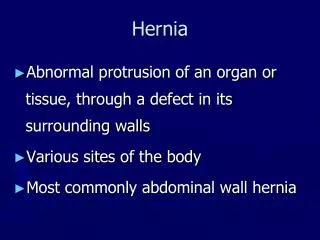
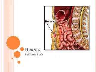
Definition A hernia is a protrusion of a viscus or part of a viscus through an abnormal opening in the walls of its containing cavity .
Anatomy • The inguinal canal :- The inguinal canal is approximately 4 cm long and is directed obliquely inferomedially through the inferior part of the anterolateral abdominal wall. The canal lies parallel and 2-4 cm superior to the medial half of the inguinal ligament.This ligament extends from the anterior superior iliac spine to the pubic tubercle. • The inguinal canal has openings at either end : – The deep (internal) inguinal ring is the entrance to the inguinal canal. It is thesite of an outpouching of the transversalis fascia. This is approximately 1.25 cm superior to the middle of the inguinal ligament The superficial, or external inguinal ring is the exit from the inguinal canal. It is a slitlke opening between the diagonal fibres of the aponeurosis of the external oblique
Inguinal canal • walls of The inguinal canal :- • The anterior wall is formed mainly by the aponeurosis of the external Oblique • . The posterior wall is formed mainly by transversalis fascia • The roof is formed by the arching fibres of the internal oblique and • transverse abdominal muscles. • The floor is formed by the inguinal ligament, which forms a shallow trough. It is reinforced in its most medial part by the lacunar ligament.
Content :- • Spermatic cord ( round ligament of the uterus in female ) The Cord Itself.—The contents of the spermatic cord are (a) the ductus (vas) deferens and its artery . (b) the testicular artery and venous (pampiniform) plexus. (c) the genital branch of the genitofemoral nerve. (d) lymphatic vessels and sympathetic nerve fibers. (e) fat and connective tissue surrounding the cord and its coverings in various amounts • Ilioinguinal nerve . • Ilioinguinal lymph node .
Femoral Canal The major feature of the femoral canal is the femoral sheath. This sheath is a condensation of the deep fascia (fascia lata) of the thigh and contains, from lateral to medial, the femoral artery, femoral vein, and femoral canal. The femoral canal is a space medial to the vein that allows for venous expansion and contains a lymph node (node of Cloquet). Other features of the femoral triangle include the femoral nerve, which lies lateral to the sheath, • Wall of The Femoral canal anterior is the inguinal ligament posterior is the iliopsoas, pectineal, and long adductor muscles (floor). Medial is lacunar ligament Lateral is femoral vessle
Predisposing: All hernias occur at the site of WEAKNESS OF THE ABDOMINAL WALL which are acted on by repeated INCREASE in abdominal pressure
repeated INCREASE in abdominal pressure is usually due to • Chronic cough • Straining • Bladder neck or urethral obstruction • Pregnancy • Vomiting • Sever muscular effort • Ascetic fluid
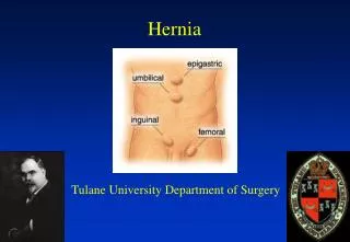
Types • Inguinal • Femoral • Epigastric • Para umbilical • Umbilical • Obturator • Superior lumbar • Inferioer lumbar • Gluteal • Sciatic • Incisional
Indirect Inguinal Hernia Hernia through the inguinal canal • Direct Inguinal Hernia The sac passes through a weakness or defect of the transversalis fascia in the posterior wall of the inguinal canal • Femoral Hernia Hernia medial to femoral vessels under inguinal ligament • Umbilical Hernia Hernia through the umbilical ring • Paraumbilical Hernia A protrusion through the linea alba just above or sometimes just below the umbilicus • Epigastric Hernia Protrusion of extraperitoneal fat through the linea alba anywhere between the xiphoid process and the umbilicus • Incisional Hernia Hernia through an incisional site • Lumber Hernia occur through the inferior lumber triangle of Petit
Inguinal hernia • History: • Age ( young vs. old) • Occupation ( nature ?? ) • Local symptoms: Swelling, discomfort and pain • Systemic symptoms: if there is obstruction or strangulation • Precipitating factors
Inguinal hernia • Examination: • Inspection for site, size, shape and color. • Palpation for surface, temp, tenderness, composition and reducibility. • Expansible cough impulse. • General exam: for common causes of increase intra abdominal pressure
Indirect Versus Direct inguinal hernias • Indirect is the most common form of hernia and its usually congenital due to patent processus viginalis • Direct usually acquired occur in old men with weak abdominal muscles.
Differential diagnosis of inguinal hernias • Male: 1 ) Femoral hernia 2 ) Vaginal hydrocele 3 ) Spermatocele 4 ) Encysted hydrocele of the cord 5 ) Un-descended testis 6 ) Lipoma of the cord • Female 1 ) Hydrocele of the canal of nuck: Is a fluid filled distal part of the sac of an indirct hernia with narrow proximal part it present with a smoth fluctuant swelling with out a cough impulse which will transilluminate 2 ) Femoral hernia Note that examination using finger and thumb across the neck of the scrotum will help to distinguish a swelling of inguinal origin and one that is entirely intrascrotal
Femoral hernia Small femoral hernia may be unnoticed by the patient or disregarded for years perhaps until the day it strangulates. Adherence of the greater omentum sometimes causes a dragging pain. Rarely a large sac is present .
Femoral hernia History • Age ; uncommon in children , most common in old age female . • Sex; women > men (but still commonest hernia in women the inguinal hernia ) • The patient came with local symptoms • 1- discomfort and pain • 2- swelling in the groin • General ; femoral hernia is more likely to be strangulated than the inguinal hernia • Multiplicity ; often bilateral
Differential diagnosis of femoral hernia 1) Inguinal hernia 2 ) saphenavarix: a saccularenlargment of the termination of the long saphenous vein The swelling disappears completely when the patient lies flat there is impulse in coughing and fluid thrill and sometimes venous hum can be heard over a saphenavarix 3 ) Enlarge lymph node: fever + other lymph node enlargment 4 ) Lipoma 5 ) Femoral aneurysm: expansile pulsation 6 ) Psoas abscess: There is often a fluctuating swelling and examination of the spine and a radiograph will confirm the diagnosis 7 ) A distended psoas bursa: The swelling diminishes when the hip is flexed and osteoarthritis of the hip is present
Umbilical hernia • Signs and symptoms • Age ; doesn’t appear until the umbilical cord has separated and healed . • No specific symptoms • Have wide neck and reduce easily , rarely give intestinal obstruction. • Nature history ; 90 % disappear spontaneously during the first year.
Examination • Inspection • Site ; in the center of the umbilicus • Size and shape ; size can vary from vary small to very large . Shape is usually hemispherical. • Palpation • Composition ; contain bowel , which makes it resonant to percussion . They reduce spontaneously when the child lies down . • Reducibility ; easy • Cough impulse; invariably present .
Acquired umbilical hernia • Hernia through the umbilical scar , so it is a true umbilical hernia. • Not common and is usually secondary to increase intra abdominal pressure. • The most common causes • 1- pregnancy • 2- ascitis • 3- ovarian cyst • 4- fibrodis • 5- bowel distention
Incision hernia • Signs and symptoms • Previous operation or accidental trauma • Age ; all ages , but more common in old age. • Symptom ; lump ,pain ,intestinal obstruction ( distention ,colic, vomiting ,constipation , sever pain in the lump ) • Examination • 1- reducible lump • 2- expansile cough impulse • 3- if the lump dose not reduse and dose not have cough impulse , than it may be not a hernia • Ddx • Tumor • Chronic abscess • Hematoma • Foreign body granuloma
Preoperative assessment • proper history and examination • identify high risk patients • prepare the preoperative notes : • consent.. • pre op Dx • procedure planned • surgeons • Anasthesia anticipated (general , local, spinal)
Preoperative assessment • Investigation data ( pre operative tests ) : 1. Lab : * CBC : to check hemoglobin level anemia and WBCs infections * U&E : to check for any electrolyte imbalance * LFTs : indicated in jaundiced patients and suspected hepatitis or any clotting problems * PT & PTT * ABG * grouping and cross matching 2. Imaging : * Chest X ray : for all patients 3. ECG : for any patient who is more than 40 years of age
Preoperative assessment • current medications or allergies • any major (chronic) illness • pre op orders : • skin preparation • diet (NPO) • GIT preparation • Sedation • Preanesthetic medications • Other medications • Antibiotics • Blood transfusion ( if needed ) • Bladder preparation
Pre op evaluation &preparation Surgical TTT Watchful Waiting May be appropriate for pt with asymptomatic hernia or elderly pt with minimal symptoms or easily reduced inguinal hernia. Routine F/U with health care professional A Randomized trial concluded that this is an acceptable option for men with minimally symptomatic inguinal hernia and that delaying repair until symptoms increase is safe due to low rate ofincarceration. 23% of pt initially treated with watchful waiting crossed over to surgical ttt due to increase in symptoms (most often hernia-related pain) , only 1 pt (0.3%) experienced acute hernia incarceration without strangulation within 2years, a second had acute incarceration with Bowel obstruction at 4 years, corresponding to frequency of acute intervention of 1.8/1000 pt-years (JAMA 2006,295:285)
Pre op preparation • Most pt are treated surgically • Increase IAP abnormalities (Chronic cough, Constipation, Bladder outlet obstruction) should be evaluated and remedied to extent possible before elective herniorrhaphy. • In case of intestinal obstruction and possible strangulation, Broad spectrum AB,NG suction may be indicated, correction of volume status& elctroyles.
Reduction • Uncomplicated: • Manual Gentle pressure over hernia Gentle traction over the mass sedation and trendelenburg position. • Complicated (strangulated): • no attempt should be made to reduce the hernia because of potential reduction of gangrenous segment of bowel with the hernial sac.
Surgerical TTT • 1.choice of anesthetic: • elective open repair : Local is preferred • Laproscopic hernia repair: more commonly under GA.
2.TTT OF HERNIAL SAC • INDIRECT: sac is dissected free from the cord structures and creamsteric fibers. Sac should be open away from any herniated contents. Contents are then reduced, and the sac is ligated deep to inguinal ring with an absorbable suture • DIRECT: • Too broadly based for ligation and should not be opened, simple freed from transversalis fibers and inverted.
3.Inguinal Floor Reconstruction • Some method of reconstruction of the inguinal floor is necessary in all adult hernia repairs to prevent recurrence.
1.Primary tissue repair • Bassini repair: inferior arch of transversalis fascia (TF) or conjoint tendon is approximated to shelving portion of inguinal ligament. • McVay: TF is sutured to cooper ligament. • Shouldice: TF is incised and reapproximated.
2.Open tension free repair • Lichtenstein repair &Patch and Plug technique: Mesh is used to reconstruct inguinal floor • Mesh plug technique : place mesh in the hernial defect
Laproscopic & preperitoneal repairs • TAPP (transabdominal prepeitoneal procedure): peritoneal space entered by conventional lap at umbilicus and peritoneum overlaying inguinal floor is dissected away as flap. • TEP (Total extraperitoneal repair): preperitoneal space is developed with a balloon inserted between posterior rectus sheath and peritoneum balloon inflated to dissect the peritoneal flaps awau from posterior abdomianl wall and the direct and indirect spaces, other ports inserted into this preperitoneal space without entering peritoneal cavity. • After lap. Dissection and reduction of hernia sac , a large piece of mesh is placed over inguinal floor
Femoral hernia repair Femoral hernias should be repaired very soon after the diagnosis has been made because of the high risk of strangulation. There is no place for a truss for a femoral hernia. Different approaches : Open VS Laparoscopic
Open surgery Three approaches have been described for open surgery : • Infra-inguinal approach (Lookwood) • Supra-inguinal approach ( McEvedy) • Trans-inguinal approach ( Lotheissen)
Each technique has the principle of dissection of the sac with reduction of its contents, followed by ligation of the sac and closure between the inguinal and pectineal ligaments.
Lockwood’s infra-inguinal approach • The sac is dissected out below the inguinal ligament via groin crease incision. • Then the sac is opened and the contents are inspected and reduced into the abdomen. • Then the neck of the sac is pulled down , ligated and allowed to retract through femoral canal. • Then close the femoral canal by mesh plug or non absorbable sutures.
McEvedy’s high approach • Vertical incision is made over the femoral canal and continued upwards above the inguinal ligament. • This incision provides good access to the preperitoneal space and then to the peritoneum itself. • Use finger dissection to sweep peritoneum from anterior abdominal wall , so the neck of the sac can be identified. • Dissect the sac , reduce the contents and repair the defect by mesh or sutures.
Lotheissen‘s trans-inguinal approach • The incision is made superior and parallel to inguinal ligament extending from pubic tubercle to mid inguinal point.