Download
1 / 39
430 likes | 848 Views
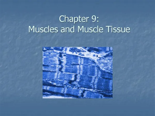
Anatomy of Muscles and Muscle Tissue. 9. Muscle Overview. The three types of muscle tissue are skeletal , cardiac , and smooth These types differ in structure, location, function, and means of activation. Muscle Similarities.

E N D
Muscle Overview • The three types of muscle tissue are skeletal, cardiac, and smooth • These types differ in structure, location, function, and means of activation
Muscle Similarities Skeletal and smooth muscle cells are elongated and are called muscle fibers Muscle contraction depends on two kinds of myofilaments – actin and myosin Muscle terminology is similar: • Sarcolemma – muscle plasma membrane • Sarcoplasm – cytoplasm of a muscle cell • Prefixes – myo, mys, and sarco all refer to muscle
Skeletal Muscle Tissue • Packaged in skeletal muscles that attach to and cover the bony skeleton • Has obvious stripes called striations • Is controlled voluntarily (i.e., by conscious control) • Contracts very rapidly, but tires easily • Is responsible for overall body motility
Cardiac Muscle Tissue • Occurs only in the heart • Is striated like skeletal muscle but is involuntary(controlled by the autonomic nervous system) • Contracts at a fairly steady rate set by the heart’s pacemaker cells • Neural controls allow the heart to respond to changes in bodily needs
Cardiac Muscle Cells A. General Features 1. involuntary muscle 2. one, centrally located nucleus 3. mitochondria larger and more numerous B. Structure of Tissue 1. muscle fibers branch and interconnect 2. intercalated disc - thickening of sarcolemma 3. cells connected by gap junctions a. allow passage of ions like Calcium b. makes adjacent cells electrically linked c. allows for rhythmic, domino-like contraction
Smooth Muscle Tissue • Found in the walls of hollow visceral organs, such as the stomach, urinary bladder, and respiratory passages • Forces food and other substances through internal body channels • It is not striated and is involuntary (controlled by the autonomic nervous system)
Smooth Muscle • Composed of spindle-shaped fibers with a diameter of 2-10 m and lengths of several hundred m • Lack the coarse connective tissue sheaths of skeletal muscle, but have fine endomysium • Organized into two layers (longitudinal and circular) of closely apposed fibers • Found in walls of hollow organs (except the heart) • Peristalsis – alternating contractions and relaxations of smooth muscles that mix and squeeze substances through the lumen of hollow organs
Smooth Muscle Figure 9.24
Smooth Muscle Cells A. General Features 1. non-striated 2. 5-10 um in diameter; 30-200 um long 3. thick in middle; thinner, tapering off to the end 4. single oval centrally located nucleus 5. actin and myosin fibers not arranged as sarcomere 6. lack of organization (no bands) --> smooth muscle 7. also contain intermediate filaments
Smooth Muscle Cells B. Difference in Contraction 1. intermediate filaments attach to dense bodies 2. as muscle cell contracts, twists like corkscrew 3. caveolae - like T tubules of skeletal muscle
Smooth Muscle Cells C. Two Kinds of Smooth Muscle 1. visceral (single unit) muscle a. small arteries and veins b. viscera - stomach, intestines, uterus, bladder c. continuous network with gap junctions d. action spreads from one cell to another 2. multiunit muscle a. each fiber (cell) has it own nerve ending b. no gap junctions c. large arteries, large airways, arrector pili
Skeletal Muscle: Nerve and Blood Supply • Each muscle is served by one nerve, an artery, and one or more veins • Each skeletal muscle fiber is supplied with a nerve ending that controls contraction • Contracting fibers require continuous delivery of oxygen and nutrients via arteries • Wastes must be removed via veins
Skeletal Muscle Each muscle is a discrete organ composed of muscle tissue, blood vessels, nerve fibers, and connective tissue The three connective tissue sheaths are: • Endomysium – fine sheath of connective tissue composed of reticular fibers surrounding each muscle fiber • Perimysium – fibrous connective tissue that surrounds groups of muscle fibers called fascicles • Epimysium – an overcoat of dense regular connective tissue that surrounds the entire muscle
One Skeletal Muscle in Cross Section Figure 9.2 (a)
Myofibrils • Myofibrils are densely packed, rodlike contractile elements • They make up most of the muscle volume • The arrangement of myofibrils within a fiber is such that a perfectly aligned repeating series of dark A bands and light I bands is evident
Many Myofibrils in One Muscle Cell Figure 9.3 (b)
Sarcomeres The smallest contractile unit of a muscle The region of a myofibril between two successive Z discs • Myofilaments are of two types – thick and thin
Many Sarcomeres make up a single Myofibril Figure 9.3 (c)
Myofilaments: Banding Pattern • Thick filaments (myosin filaments) – extend the entire length of an A band • Thin filaments (actin filaments) – extend across the I band and partway into the A band
Myofilaments: Banding Pattern Figure 9.3 (c, d)
Ultrastructure of Myofilaments: Thick Filaments • Thick filaments are composed of the protein myosin • Each myosin molecule has a rodlike tail and two globular heads • Tails – two interwoven, heavy polypeptide chains • Heads – two smaller, light polypeptide chains called cross bridges
Ultrastructure of Myofilaments: Thick Filaments Figure 9.4 (a)(b)
Ultrastructure of Myofilaments: Thin Filaments • Thin filaments are composed of the protein actin • Each actin molecule is a helical polymer of globular subunits called G actin • The subunits contain the active sites to which myosin heads attach during contraction • Tropomyosin and troponin are regulatory subunits bound to actin
Ultrastructure of Myofilaments: Thin Filaments Figure 9.4 (c)
Arrangement of the Filaments in a Sarcomere • Longitudinal section within one sarcomere Figure 9.4 (d)
Sarcoplasmic Reticulum (SR) • SR is an elaborate, smooth endoplasmic reticulum that mostly runs longitudinally and surrounds each myofibril • Functions in the regulation of intracellular calcium levels • T tubules associate with the paired terminal cisternae to form triads
Sarcoplasmic Reticulum (SR) Figure 9.5
Neuromuscular Junction (NMJ) 1. motor neuron - nerve cell that innervates muscle 2. motor end plate - where axon meets muscle cell 3. neuromuscular junction - entire muscle/nerve site 4. synapse – name for where nerve terminal meets target cell 5. synaptic vesicles - inclusions in the axon terminal a. neurotransmitter - chemical messenger i. acetylcholine ACh (for muscular synapse) 6. synaptic cleft - space between axon and cell 7. motor unit - a neuron and all muscle cells it stimulates
Muscle Hypertrophy and Atrophy 1. muscular hypertrophy a. increase in size of myofibers (muscle cells) b. allows for increased strength c. adult muscle cells are amitotic (do not divide) 2. muscular atrophy a. progressive loss of myofibrils i. disuse atrophy ii. denervation atrophy (neuron lost)
Interactions of Skeletal Muscles • Skeletal muscles work together or in opposition • Muscles only pull (never push) • As muscles shorten, the insertion generally moves toward the origin • Whatever a muscle (or group of muscles) does, another muscle (or group) “undoes”
Muscle Classification: Functional Groups Prime movers – provide the major force for producing a specific movement Antagonists – oppose or reverse a particular movement Synergists– work cooperatively to achieve a certain action • Add force to a movement • Reduce undesirable or unnecessary movement Fixators – synergists that immobilize a bone or muscle’s origin
Naming Skeletal Muscles 1. Location of muscle – bone or body region associated with the muscle 2. Shape of muscle – e.g., the deltoid muscle (deltoid = triangle) 3. Relative size – e.g., maximus (largest), minimus (smallest), longus (long) 4. Direction of fibers – e.g., rectus (fibers run straight), transversus, and oblique (fibers run at angles to an imaginary defined axis)
Naming Skeletal Muscles 5. Number of origins – e.g., biceps (two origins) and triceps (three origins) 6. Location of attachments – named according to point of origin or insertion 7. Action – e.g., flexor or extensor, as in the names of muscles that flex or extend, respectively