Download
1 / 1
10 likes | 132 Views
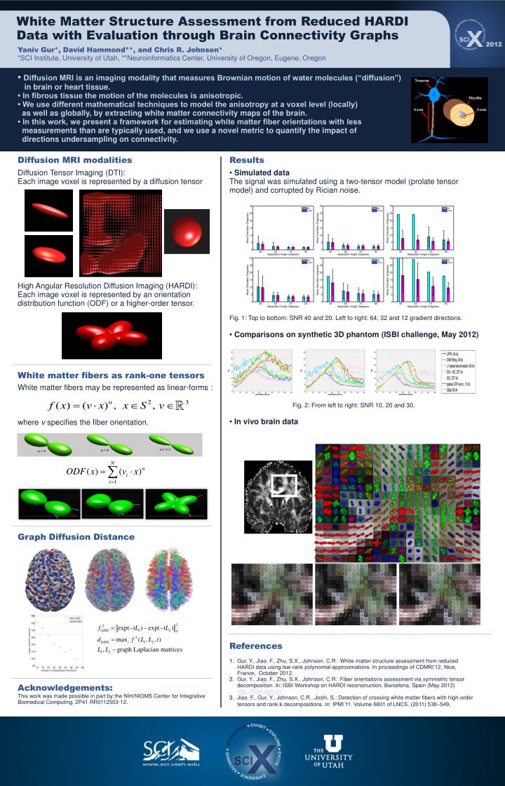
White Matter Structure Assessment from Reduced HARDI Data with Evaluation through Brain Connectivity Graphs. Yaniv Gur*, David Hammond**, and Chris R. Johnson* *SCI Institute, University of Utah, **Neuroinformatics Center, University of Oregon, Eugene, Oregon.

E N D
White Matter Structure Assessment from Reduced HARDI Data with Evaluation through Brain Connectivity Graphs Yaniv Gur*, David Hammond**, and Chris R. Johnson* *SCI Institute, University of Utah, **Neuroinformatics Center, University of Oregon, Eugene, Oregon • Diffusion MRI is an imaging modality that measures Brownian motion of water molecules (“diffusion”) in brain or heart tissue. • In fibrous tissue the motion of the molecules is anisotropic. • We use different mathematical techniques to model the anisotropy at a voxel level (locally) as well as globally, by extracting white matter connectivity maps of the brain. • In this work, we present a framework for estimating white matter fiber orientations with less measurements than are typically used, and we use a novel metric to quantify the impact of directions undersampling on connectivity. Diffusion MRI modalities Results Diffusion Tensor Imaging (DTI): Each image voxel is represented by a diffusion tensor High Angular Resolution Diffusion Imaging (HARDI): Each image voxel is represented by an orientation distribution function (ODF) or a higher-order tensor. • Simulated data • The signal was simulated using a two-tensor model (prolate tensor model) and corrupted by Rician noise. • Fig. 1: Top to bottom: SNR 40 and 20. Left to right: 64, 32 and 12 gradient directions. • Comparisons on synthetic 3D phantom (ISBI challenge, May 2012) • In vivo brain data Fig. 2. Comparisons with multi-tensor fitting. Top to bottom: SNR 40 and 20. Left to right: 64, 32 and 12 gradient directions. White matter fibers as rank-one tensors White matter fibers may be represented as linear-forms : where v specifies the fiber orientation. Fig. 2: From left to right: SNR 10, 20 and 30. Graph Diffusion Distance References Gur, Y., Jiao, F., Zhu, S.X., Johnson, C.R.: White matter structure assessment from reduced HARDI data using low-rank polynomial approximations. In proceedings of CDMRI’12, Nice, France, October 2012. Gur, Y., Jiao, F., Zhu, S.X., Johnson, C.R.: Fiber orientations assessment via symmetric tensor decomposition. In: ISBI Workshop on HARDI reconstruction, Barcelona, Spain (May 2012) http://hardi.epfl.ch/ Jiao, F., Gur, Y., Johnson, C.R., Joshi, S.: Detection of crossing white matter fibers with high-order tensors and rank-k decompositions. In: IPMI’11. Volume 6801 of LNCS. (2011) 538–549. Acknowledgements: This work was made possible in part by the NIH/NIGMS Center for Integrative Biomedical Computing, 2P41 RR0112553-12.