Download
1 / 26
280 likes | 662 Views
Development of Animals . Aka…Embryology. Female Reproductive System. Embyro Diagram. Embryology - study of development of the embryo. 5 major stages.. 1. Gametogenesis - gamete production 2. Fertilization - gamete --> zygote 3. Cleavage - Zygote --> Blastula

E N D
Development of Animals Aka…Embryology
Embryology - study of development of theembryo 5 major stages.. • 1. Gametogenesis - gamete production • 2. Fertilization - gamete --> zygote • 3. Cleavage - Zygote --> Blastula • 4. Gastrulation - Blastula --> Gastrula • 5. Organogenesis - Organ Formation • -i.e. Neurulation- Gastrula --> Neurula
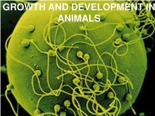
A. Fertilization:male and female gametes fuse toform a zygote 1. Funnel-shaped end of a fallopiantube is the usual site of fertilization 2. If a sperm penetrates the egg, fertilization results. Tiny hair-like cilia lining fallopian tube propel fertilized egg, zygote, through the tube toward the uterus.
Cell Division1…2 buckle my shoe • Cleavage: process by which a zygote divides by mitosis to form two new cells a. Two cells will divide to form 4 cells, and so on b. Continue dividing until a blastula (a.k.a. blastocyst) is formed
4 cell stage • 2 cells divide by mitosis and become 4 • 2nd mitotic division • Still the same size as the previous cleavage (2 cell)
8 cell • 1st time it’s obvious that the cell is either a human or another organism like a worm! • You can tell it is a deuterostome or a protostome….. What’s that??
Deuterostomes vs. ProtostomesSAY WHAT??? • 8-CELL STAGE IS KEY DIFFERENCE!!!-Deuterostomes- (starfish & vertebrates) cleavage results in 8 cells sitting directly on top of each other. This is called radial cleavage
Protostomes • Protostomes (clams, worms, & insects) undergo spiral cleavage- cells divide and they do not sit on top of each other; they appear to spiral
2. MORULA – 16 cell 2. Morula: a zygote consisting of about 12-32 cells in a solid ball that reaches uterus in about 3-4 days after fertilization
3. Blastula (blastocyst) formation 3. Blastula= a hollow ball of cells that forms between 5-8 days after fertilization • This will attach to the lining of the uterus • cell division continues Cells start to compact and move to the edge of the cell, leaving a fluid-filled space in the center- which is called the blastocoel
4. Gastrula • Now gastrulation occurs. The hollow ball of cells known as the blastula begins to fold inward on one side • (imagine that you had a deflated basketball and pushed it in on one side). • When the folding occurs, it begins to create a horse shoe shaped structure that is 2 cell layers thick..
GastrulationBlastula (hollow ball of cells) transformed into the Gastrula (three-layered stage) • Gastrula: structure made up of 2 layers of cells with an opening at one end • The point where the horse shoe almost touches is called the blastopore (small hole). • In protostomes this will eventually form the mouth. • In deuterostomes this will form the anus. • Cells that are folding inward form a cavity lined with a 2nd layer of cells • Gastrulation - sorts all the cells into distinct cell layers (ectoderm, endoderm, and mesoderm)
Gastrulation forms Three distinct tissue layers a. Layer on the outer surface of the gastrula is called the ECTODERM • 1. ECTODERM Continues to divide and eventually forms the SKIN and NERVOUS tissue
b. ENDODERM • B. Layer on the INNER SURFACE of the gastrula is called the ENDODERM • 1. ENDODERM Continues to divide and eventually develops into the lining of the digestive tract and organs associated with digestion
c. MESODERM 1.Mesoderm is the 3rd cell layer found in the developing embryo between the ECTO and ENDODERM (“meso” means middle) 2. MESODERM Continues to grow and divide and eventually develops into muscle cells, circulatory, and excretory cells, bone cells and respiratory system