Download
1 / 55
570 likes | 643 Views
Rhinosinusitis. Prof. Dr. Yavuz Selim Pata. Introduction. Sinusitis diagnosis rare 25 years ago Better understanding pathophysiology, etiology, treatment outcomes Better diagnostic techniques 5-10% of viral URI’s complicated by bacterial rhinosinusitis

E N D
Rhinosinusitis Prof. Dr. Yavuz Selim Pata
Introduction • Sinusitis diagnosis rare 25 years ago • Better understanding • pathophysiology, etiology, treatment outcomes • Better diagnostic techniques • 5-10% of viral URI’s complicated by bacterial rhinosinusitis • Numerous controversies in diagnosis and treatment
Anatomy • Maxillary Sinus • first to develop at day 65 of gestation • seen on plain films at 4-5 months • growth in phases at 3 years and 7 to 12 years • slow expansion until 18 years • average capacity is 14.75 mL • drains into middle meatus
Anatomy • Ethmoid Sinus • develop in third month of gestation • anterior from the lateral nasal wall • posterior from superior meatus • ethmoids seen on radiographs at one year • enlarges to reach adult size at age 12 • 4-17 cells each side with volume 15 mL • drainage into middle and superior meatus
Anatomy • Frontal Sinus • begins in fourth month of gestation from superior ethmoid cells • seen on radiographs at age 5-6 • grows slowly to adult size by adolescence • volume of 5-6 mL with variable development • drains into frontal recess
Anatomy • Sphenoid Sinus • originates in fourth gestational month from posterior part of nasal cavity • pneumatization begins at age 3 • rapid growth to reach sella by age 7 and adult size at age 18 • volume of 7.5 mL with drainage into superior meatus
Histology • Pseudostratified columnar epithelium • Cilia specifically arranged • Similar mucosa to remainder of tracheobronchial tree
Pathophysiology and Etiology • Normal function • patent ostia • normal cilia • normal mucous secretions • Primary sinus abnormality is obstruction of the osteomeatal complex by edema or mechanical obstruction
Etiology • Obstruction leads to retained secretions resulting in hypoxia of sinus mucosa--causes ciliary dysfunction and increased secretions-- secondarily infected • Edema and mechanical obstruction • local factors • regional factors • systemic factors • others
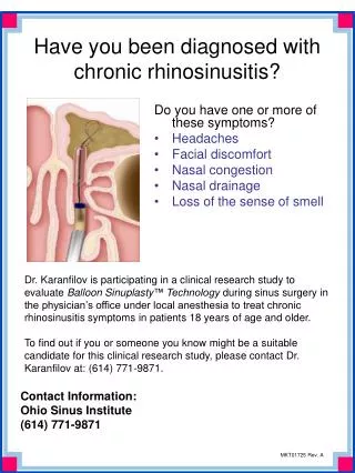
Definitions • Rhinosinusitis • unable to differentiate clinically • isolated sinusitis rare • Acute Rhinosinusitis • infection that resolves within 12 weeks • no URI during this 3 month period • divided into severe and nonsevere forms
Definitions • Recurrent Acute Rhinosinusitis • repeated acute episodes completely resolving within 12 week time frame • Chronic Rhinosinusitis • low grade symptoms and signs persistent for over 12 weeks • acute exacerbations can occur
Clinical Presentation • History and PE vital to proper diagnosis • Viral URI • unable to differentiate within 10 days • serous rhinorrhea--may be mucopurulent • nasal congestion and cough prominent • low grade fevers, malaise, headaches • nighttime cough may linger
Clinical Presentation • Acute Nonsevere Rhinosinusitis • persistent cold symptoms over 10 days • rhinorrhea (any type), cough (dry or wet) worse at night, low grade fevers, fetid breath, painless periorbital swelling in AM, rarely facial pain
Clinical Presentation • Acute Severe Rhinosinusitis • usually after 10 days but may be sooner • high fever, purulent and copious rhinorrhea, periorbital swelling, facial pain, headaches, dental pain
Diagnosis • History • Physical Examination • anterior rhinoscopy with otoscope • oropharynx • tenderness over sinuses • periorbital edema and discoloration • flexible and rigid endoscopy in older child • most specific-- mucopurulence, periorbital swelling, facial tenderness
Diagnosis • Transillumination -- no value • Ultrasonography -- little value • Radiography • traditional views Water’s, Caldwell, Lateral, and Submentovertex • problems: ethmoids, disease findings, underdeveloped sinuses
Diagnosis • Radiography • McAlister: compared radiographs with CT -- 45% normal X-ray but abnormal CT 34% abnormal x-ray but normal CT • Not useful for uncomplicated rhinosinusitis • Uses in complicated acute rhinosinusitis • with AFL -- 75% positive isolates
Diagnosis • Computed tomography • gold standard • planning surgery or failed medical management • Indications • Clinical unresponsiveness to medical therapy • Immunosuppressed patient • Severe symptoms or signs • Life threatening complications
Diagnosis • Sinus Aspirate • indications same for CT scanning • nasal, oral, nasopharyngeal cultures poor • needs cooperative patient -- usually GETA • middle meatal cultures?
Microbiology • Similar to adults • Streptococcus pneumoniae, Moraxella catarralis, nontypeable Hemophilus influenzae • Rare viruses, anaerobes, Staphylococcus • Normal flora in the sinus-- controversy
Medical Management • Historically -- aspiration and irrigation • Antibiotics -- viral URI common and increasing numbers of drug resistant bacteria • 40-60% sinusitis episodes resolve (AOM) • 35% of S. pneumoniae penicillin-resistant • 16% of S. pneumoniae penicillin-intermediate • rapid cure, prevent complications, prevent chronic sinusitis, sterilize sinus
Medical Management • Acute Nonsevere Rhinosinusitis (no ABX) • Amoxicillin (45-90 mg/kg/day), amoxicillin/clavulanate, cefpodoxime, or cefuroxime • 10 to 14 day course • PCN-allergic may receive azithromycin, clarithromycin, erythromycin, or TMP/SMX but limited effectiveness (25% failure rate)
Medical Management • Acute nonsevere rhinosinusitis (with ABX) Acute severe rhinosinusitis (no ABX) • Amoxicillin/clavulanate, high dose amoxicillin (80-90 mg/kg/day), cefpodoxime, or cefuroxime • Acute severe rhinosinusitis (with ABX) • amoxicillin/clavulanate or combination therapy (amoxicillin or clindamycin plus cefpodoxime or cefixime)
Medical Management • Complications or severe illness • IV cefotaxime or ceftriaxone plus clindamycin • Chronic Rhinosinusitis • beta lactam stable agent (amoxicillin/clavulanate or combination therapy) for 3-6 weeks
Medical Management • Antihistamines -- dry mucosal secretions • Isotonic saline nose drops, sprays, irrigations, and steam inhalations -- anecdotal • Topical decongestants --inhibits cilial motion • Nasal steroids • Mucolytics
Medical management • Recalcitrant rhinosinusitis • allergy • immunodeficiency • cystic fibrosis • ciliary dismotility disorders • gastroesophageal reflux disease
Surgical Management • Adenoidectomy • nasal obstruction and symptoms • small size of trials • Septoplasty • rare to have significant septal deviation • Antral aspiration and lavage • indications same as sinus aspiration • only treats maxillary sinus • need GETA
Surgical Management • Caldwell-Luc -- damages dentition • Inferior antrostomy • goes against proven cilial outflow • possible for cilial dismotility/CF • FESS • controversial -- difficult, too radical (AOM), reversible changes on CT
Surgical Management • FESS • excellent results : 71% normal at one year, meta analysis 89% success with 0.6% complications • usually maxillary antrostomy/anterior ethmoidectomy
Surgical Management • FESS (absolute) • complete nasal obstruction in CF • antrochoanal polyp • intracranial or orbital complications • mucocoeles or mucopyocoeles • traumatic injury in optic canal • resistant dacryocystorhinitis • fungal sinusitis • some meningoencephaloceles/neoplasms
Surgical Management • FESS (possible) • persistent chronic rhinosinusitis that fails optimum medical treatment and after exclusion of systemic disease • asthmatic exacerbations associated with rhinosinusitis
Complications • Routes of spread • arterial • venous • lymphatic • direct
Complications • Stage I • periorbital inflammatory edema • obstruction of venous channels • no vision loss • no EOM limitation
Complications • Stage II • orbital cellulitis with edema, chemosis, proptosis, pain • no abscess • opthalmoplegia may occur due to edema or spasm • no visual loss
Complications • Stage III • subperiosteal abscess • globe displaced laterally or downward • orbital cellulitis present with decreased EOM • vision decreased
Complications • Stage IV • orbital abscess • severe proptosis and chemosis • usually no globe displacement • opthalmoplegia present • visual loss (13%) due to ischemia or neuritis
Complications • Stage V • cavernous sinus thrombosis • progressive symptoms • proptosis and fixation • CN II, IV, VI • meningitis • high mortality
Complications • History an physical examination • Ophthalmology consultation • IV antibiotics (ceftriaxone plus metronidazole and oxacillin) • CT scan • Surgery -- abscess, worsening vision, progression, persistent after 24 hours • external, FESS, frontal sinus trephine
Complications • Intracranial --meningitis, subdural or epidural abscess, cerebral abscess, CST • neurosurgery, ophthalmology, ID
Allergy and Rhinosinusitis • Allergy estimated at 15-30% of population • Major contributing factor in rhinosinusitis • Similar pathogenesis as viral etiology with obstruction -- mucostasis --hypoxia -- colonization
Allergy Diagnosis • History is critical • itching mucous membranes, clear rhinorrhea, eczema, food intolerance, nasal congestion, stuffiness, fluctuating rhinorrhea, sneezing, cough, behavioral changes, headaches, facial pressure • prior history of infantile colic, formula changes, otitis media, ADHD
Allergy Diagnosis • Physical Examination • allergic shiners and allergic salute • nasal obstruction with cracked lips • rash over cheeks or urticaria • eczema • posterior pharyngeal lymphoid tissue • ETD
Allergy Diagnosis • Clinical diagnosis • Two to four week food diary • Open feeding challenge • RAST testing -- poor for food allergy • Nasal smear analysis • Skin testing