Download
1 / 52
830 likes | 1.9k Views
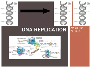
DNA Replication. The basic rules for DNA replication. DNA Polymerases. Initiation of replication. DNA synthesis at the replication fork. Termination of replication. Regulation of re-initiation. Other modes of DNA replication. Termination of replication.

E N D
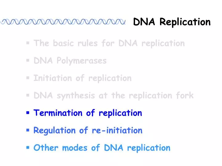
DNA Replication The basic rules for DNA replication DNA Polymerases Initiation of replication DNA synthesis at the replication fork Termination of replication Regulation of re-initiation Other modes of DNA replication
Termination of replication Circular bacterial chromosomes Linear chromosomes
Forks meet For the circular bacterial replicon, the two replication forks move around the genome to a meeting point. Replication fork Replication fork
oriC E. coli K-12 4,639,221 bp Terminus 圖引用自:Nelson, D. L. and Cox, M. M. (2005) Lehninger Principles of Biochemistry. 4th Ed., Worth Publishers. Fig. 25-1
ter sites: E, D, A C, B Binding of Tus protein to a ter site arrests replication fork advancement. Figure 13.7
Tus protein terminator utilization substance • Encoded by the tus gene A 309-residue monomer 圖引用自:Voet, D., Voet, J. G. and Pratt, C.W. (1999) Fundamentals of Biochemistry. John Wiley & Sons, Inc. Fig. 24-16
Figure 14.34 Tus binds to ter asymmetrically and blocks replication in only one direction.
Type II topoisomerases are required to separate daughter DNA molecules. Nelson, D. L. and Cox, M. M. (2005) Lehninger Principles of Biochemistry. 4th Ed., Worth Publishers. Fig. 25-17b
Catenanes (Type II topoisomerase) Nelson, D. L. and Cox, M. M. (2005) Lehninger Principles of Biochemistry. 4th Ed., Worth Publishers. Fig. 25-17b
The ends of linear DNA are a problem for replication. Linear bacterial chromosome 3’ 5’ 5’ 3’ 5’ 3’ 3’ 5’ Last Okazaki fragment 3’ 5’ 5’ 3’ + 3’ 5’ 5’ 3’ Primer removal and ligation of Okazaki fragments
3’ 5’ 5’ 3’ + 3’ 5’ 5’ 3’ Replicate again 3’ 5’ 5’ 3’ + 3’ 5’ 5’ 3’ + 3’ 5’ 5’ 3’ + 3’ 5’ 5’ 3’ The chromosome becomes shorter.
Linear eukaryotic chromosome Replication fork Replication fork
Leading strand Lagging strand 5’ 3’ 5’ 3’ 3’ 5’ 5’ 3’ Lagging strand Leading strand Replication fork Replication fork DNA polymerase cannot synthesize the extreme 5’ ends of linear DNA.
How do cells solve the end replication problem? • Using terminally attached protein to provide an OH (in certain species of bacteria) • Using telomerase to extend the ends of chromosome (in eukaryotic cells)
Telomeres: the ends of eukaryotic chromosomes Telomere Telomere Centromere
Telomeric DNA: (Tx Gy)n Tx Gy Tx Gy 3’ (Ax Cy)n x, y : 1 ~4 Tetrahymena thermophila TTGGGG n: 20 ~ 100 in single- cell eukaryotes; T(G)2-3(TG)1-6 Saccharomyces cerevisiae > 1500 in mammals Arabidopsis thaliana TTTAGGG Human TTAGGG
Mammalian telomeres end in a large duplex loop. T loop 取材自:Griffith, J. D., Comeau, L., Rosenfield, S., Stansel, R. M., Bianchi, A., Moss, H., and de Lange, T. (1999) Mammalian telomeres end in a large duplex loop. Cell, 97: 503-514, Fig. 3.
The 3’ single-stranded end of the telomere displaces the homo-logous repeats from duplex DNA to form a t-loop. The reaction is catalyzed by TRF2. Figure 19.30
Telomeric DNA is synthesized and maintained by telomerase. Telomerase: a ribonucleoproteins RNA: as a template Protein: reverse transcriptase
Telomerase uses its RNA component to anneal to the 3’ end of ssDNA region of the telomere. RNA template directs addition of nucleotides to 3’ end of DNA 1 2 3 Telomerase moves to the newly synthesized 3’end. Figure 19.31
5’ 3’ 5’ 3’ 5’ 3’ 5’ 3’ extended by telomerase 5’ 3’ 5’ 3’ DNA synthesized by DNA Pol RNA primer
Summary Termination of replication 1. For circular bacterial chromosome: • The two replication forks of E. coli chromosome initiate at oriC, move around the genome and then meet at a ter site. • Binding of Tus protein to a ter site arrests replication fork advancement. • Type II topoisomerases are required to separate catenated daughter DNA molecules.
Termination of replication Summary 2. For eukaryotic linear chromosome: • Telomerase solves the end problem by extending the 3’ end of the chromosome. • Telomerase is a reverse transcriptase and specifically elongates the 3’OH of particular ssDNA sequences using its own RNA as a template. • Type II topoisomerases are also critical to the segregation of large linear daughter chromosomes.
DNA Replication The basic rules for DNA replication DNA Polymerases Initiation of replication DNA synthesis at the replication fork Termination of replication Regulation of re-initiation Other modes of DNA replication
In bacterial cells, methylation at the origin may regulate initiation. Me (Hemimethylated) (Fully methylated) Figure 14.36
oriC contains 11 GATC repeats that are methylated on adenine on both strands. Replication generates hemimethylated DNA. The hemimethylated origins cannot initiate again until the Dam methylase has converted them into fully methylated origins. ~ 13 min delay Figure 14.35
What is responsible for controlling reuse of origins? -- Several mechanisms may be involved: Physical sequestration of the origin Regulation of methylation by SeqA Regulation of DnaA binding by membrane-associated inhibitor by DnaAATP levels by repression of DnaA transcription
In eukaryotic cells, licensing factor control the re-initiation of replication. Figure 14.39
The replicator (origin) of S. cerevisiae: ARS(Autonomously replicating sequence) Origin recognition complex (ORC) (Initiation complex) • ORC is associated with yeast origins throughout • the entire cell cycle. 圖引用自:Cooper, G. M. (1997) The cell: a molecular approach. ASM Press. Fig. 5.17
Figure 14.40 Cdc6 is rapidly degraded during S phase, preventing re-initiation. S phase When replication is initiated, Cdc6 and MCM proteins are displaced.
DNA Replication The basic rules for DNA replication DNA Polymerases Initiation of replication DNA synthesis at the replication fork Termination of replication Regulation of re-initiation Other modes of DNA replication
Chromosome in different organisms: Prokaryotes Eukaryotes
Extrachromosomal DNA: DNA in eukaryotic organelles Mitochondrial DNA Chloroplastic DNA Plasmid Viral DNA
2. Replication begins at an origin. • Replication loops always initiate at a unique point, called an origin. For most eukaryotic and prokaryotic DNAs: 1. Both DNA strands are replicated simultaneously. 2. Replication is bidirectional. Some extrachomosomal DNAs are replicated unidirectionally or using different replication modes.
Other modes of DNA replication • Replication of mitochondrial DNA Replication of adenovirus DNA • Rolling-circle replication of bacteriophage ssDNA Replication and transfer of F plasmid
Mitochondrial DNA: L strand origin H strand origin L strand H strand
H H L H H RNA primer Initiation at origin on H Synthesis of DNA (H as template) D loop Newly synthesized strand displaces L strand Based on Figure 13.11
Replication of the L-strand is initiated when its origin is exposed. H L H H L L
Replication of adenovirus DNA: DNA synthesis initiates at left 5’ end. duplex origin Single strand is displaced when fork reaches end. Figure 13.13
Terminal proteins enable initiation at the ends of adenovirus DNA. Figure 13.15 Figure 13.14
3’ 5’ • Rolling-circle replication of bacteriophage circular ssDNA: (+) DNA Pol RNA Pol (-) 3’ RNA primer 5’ Ligase (+) strand Replicative form
(+) (-) (+) (-) 3’-OH Nick at origin of (+) strand 5’-P The newly synthesized strand displaces old (+) strand. After 1 revolution displaced strand reaches unit length. (+) (+) (-) Based on Figure 13.16
(+) (+) (-) (+) (+) (-) (+) (s replication) Continued elongation generates displaced strand of multiple unit lengths
Replication of phage fX174: - A + + (+) strand Replicative form Rolling circle replication
A protein nicks the origin and binds to 5’ end. DNA replication displaces (+) strand. Replication fork passes origin, A protein nicks DNA & binds to the new 5’ end. Released (+) strand forms covalent circle. Based on Figure 13.19
F cm ~ 100 kb F cm F plasmid (F factor): An F plasmid can exist as a free circular plasmid or can integrate into the bacterial chromosome.
The F factor codes for specific pilli that form on the surface of the bacterium. An F-pilus enables an F+ bacterium to contact an F-bacterium and to initiate conjugation. Figure 13.21
Donor (F+) Recipient (F-) TraY/I nick DNA at oriT. TraY/I TraY/I multimer migrates around circle, unwinding DNA. Single strand enters recipient. Figure 13.22
Donor Recipient Complementary strands are synthesized. Donor gap is closed. Recipient circularizes. Figure 13.22
F factor is nicked at oriT. • 5’end leads ssDNA into recipient. • ssDNAs are converted to dsDNA in both bacteria. Figure 13.23