Download
1 / 43
460 likes | 1.02k Views
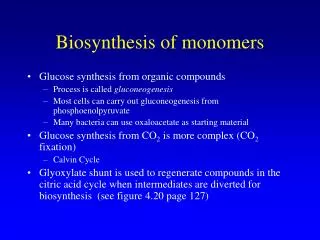
Biosynthesis of Heme. Dr.S.Chakravarty ,MD. Biosynthesis of Heme. TISSUES :- ALL TISSUES BUT PREDOMINANTLY IN THE LIVER AND BONE-MARROW SUBCELLULAR SITE :- PARTLY IN MITOCHONDRION ( FIRST STEP AND THE LAST THREE STEPS ) AND

E N D
Biosynthesis of Heme Dr.S.Chakravarty ,MD
Biosynthesis of Heme • TISSUES :- ALL TISSUES BUT PREDOMINANTLY IN THE LIVER AND BONE-MARROW • SUBCELLULAR SITE :- PARTLY IN MITOCHONDRION ( FIRST STEP AND THE LAST THREE STEPS ) AND PARTLY CYTOSOLIC. (REMAINING STEPS) • STARTING MATERIALS :- 1)Succinyl CoA( FROM TCA CYCLE) 2)AA - GLYCINE
MITOCHONDRIA 1 COMMITTED STEP & RATE –LIMITING Lead Inhibits 2 MITOCHONDRIA
FOUR MOLECUES OF PRORPHOBILINOGEN Also called PBG deaminase 3 HYDROXYMETHYLBILANE (LINEAR TETRAPYRROLE) 4 TYPE I UROPORPHYRINOGEN TYPE III UROPORPHYRINOGEN
5 ACETYL TO METHYL ( AM )
COPROPORPHYRINOGEN III 6 2CO2 PROPIONYL TO VINYL (PV ) Coporporphyrinogenoxidase PROTOPORPHYRINOGEN III 7 Protoporphyrinogen oxidase M I T O C H O N D R I A PROTOPORPHYRIN III Fe +2 8 Ferrochelatase HEME
INCORPORATION OF IRON TO PROTOPORPHYRIN Lead inhibits Iron is coordinately linked with 5N (4 Pyrrole and 1 N2 of a His residue of Globin)
UroporphyrinogenI synthase UroporphyrinogenIII synthase Uroporphyrinogendecarboxylase CYTOSOL Coporporphyrinogenoxidase 6 Protoporphyrinogen oxidase 7 MITOCHONDRIA 8 Ferrochelatase
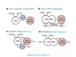
REMEMBER :- • 2/3 rd of TOTAL HEME SYNTHESIZED GOES FOR SYNTHESIS OF cytP450. • cytP450 is involved in metabolism of drugs and xenobiotics.
Regulation • The rate limiting enzyme is ALA SYNTHASE which exists in two forms ALAS1(HEPATIC ) & ALAS2 (ERYTHROID ) Feedback Regulation of ALAS1:- • HEME with the help of a APO-REPRESSOR acts as a NEGATIVE REGULATOR. • So, the Rate of synthesis of ALA SYNTHASE greatly increases in the absence of heme and is diminished in its presence.
F E E D B A C K I N H I B I T I O N
MANY DRUGS CAN CAUSE MARKEDLY INCREASE ALA SYNTHASE . -WHY ? • Several factors can cause DEREPRESSION OF ALAS-1 IN LIVER eg. • Glucose administration • Administration of HEMATIN . • ALAS-2 DOESNOT UNDERGO FEEDBACK REGULATION BY HEME . • ALAS-2 IS NOT INDUCED BY DRUGS THAT INDUCE ALAS-1.
Porphyrins are coloured and Fluoresce • The Porphyrinogens are colourless, whereas the various Porphyrins and are coloured. • There is a characteristic absorption curve for each porphyrin. For eg.( in 5%HCl ) SORET BAND Peak absorption of hematoporphyrins at 400nm
The Porphyrias • The porphyrias are a group of genetic disorders due to abnormalities in the pathway of biosynthesis of heme. • The symptoms produced depend on the type of defect. • Autosomal dominant porphyrias include acute intermittent porphyria, most cases of erythropoieticprotoporphyria, hereditary coproporphyria, and variegate porphyria. • Most common is PORPHYRIA is AIP
If the lesion occurs early in the pathway prior to the formation of porphyrinogens ALA and PBG will accumulate in body tissues and fluids . • Clinically they will complain of abdominal pain and neuropsychiatric symptoms.
Spontaneous oxidation • If the enzyme is blocked later in the pathway it will lead to accumulation of porphyrinogens . Porphyrinogens 2H Porphyrin derivatives PHOTOSENSITIVITY • The porphyrins when exposed to light of 400nm become excited and then react with molecular oxygen to produce oxygen radicals. • These radicals injure lysosomes and other cellular organelle. The lysosomes release degradative enzymes which cause various damage to skin , resulting in SCARRING and damage to organs like the Liver. Light
Acute porphyrias • Central Nervous system, • resulting in abdominal pain, vomiting, acute neuropathy, muscle weakness, seizures • mental disturbances, including hallucinations, depression, anxiety, and paranoia. • SEVERE PAIN – BOTH ACUTE AND CHRONIC • Constipation is frequently present, as the nervous system of the gut is affected, but diarrhea can also occur. • Patients with acute porphyria (AIP, HCP, VP) are at increased risk over their life for hepatocellular carcinoma (primary liver cancer) and may require monitoring. Other typical risk factors for liver cancer need not be present.
Cutaneous pophyrias • Photosensitivity and skin lesions
Gingival recession GUNTHER’S DISEASE(Congenital Erythropoieticporphyria )
GUNTHER’S DISEASE (Congenital Erythropoieticporphyria ) Erythrodontia
Why accumulation PBG and ALA lead to complications: • ALA and PBG are Neurotoxic • Vasoconstriction of CNS and abdominal blood vessels. • Decreased Heme containing proteins - Decreased cellular energy. • Elevation of stress hormones – Anxiety, Increase in BP etc.
PRECIPITATING FEATURES • DRUGS :- Certain medicines, such as BARBITURATES(SEDATIVE ) , GRISEOFULVIN (ANTI-FUNGAL)birth control pills, antibiotics, and medicines for treating seizures. • Why ? THEY ARE METABOLISED BY CYT P450 , hence UTILIZATION of HEME in CYTOCHROME P450 IS INCREASED .This DECREASES THE INTRACELLULAR CONCENTRATION. • Dehydration (loss of too much water and salt) too much sun exposure.
Cigarette smoking and alcohol consumption. • Hormone changes,during menses , menarche and menopause. • Physical or mental stress, such as with an infection, depression, emotional problem, or after surgery. • Starvation due to fasting or crash dieting.
Why accumulation PBG and ALA lead to complications: • ALA and PBG are Neurotoxic • Vasoconstriction of CNS and abdominal blood vessels. • Decreased Heme containing proteins - Decreased cellular energy. • Elevation of stress hormones – Anxiety, Increase in BP etc.
DIAGNOSIS • Porphyria is diagnosed through spectroscopy and biochemical analysis of blood, urine, and stool. • In general, urine estimation of porphobilinogen (PBG) is the first step if acute porphyria is suspected. • In nearly all cases of acute porphyria syndromes, urinary PBG is markedly elevated except for the very rare ALA dehydratase deficiency.
Diagnosis • Urinary ALA and PBG are always markedly increased in symptomatic patients with AIP and even in some asymptomatic individuals with the inherited enzyme deficiency. PBG in urine is oxidized to porphobilin upon standing, which gives a dark-brown color to urine, and often referred to as ‘port-wine reddish urine’.
Lab diagnosis: • Urinary PBG excretion during an attack : • usually 50–200 mg/24 h [normal, 0–4mg/24 h] • Urinary ALA excretion : • 20–100 mg/24 h [normal, 1–7 mg/24 h] • Portwine color of urine on prolonged standing
Repeat testing during an attack and subsequent attacks may be necessary in order to detect a porphyria, as levels may be normal or near-normal between attacks. • UV fluorescence :- Presence of porphyrins in urine is detected by Ehrlich’s reagent. If urine is observed under UV light it emits strong red-fluorescence if porphyrins are present. • DNA analysis to identify the causative mutation in the appropriate gene (AIP:HMBS; VP: PPOX; HCP: CPO ) is the method of choice. • Enzyme measurements are not as specific or sensitive as DNA analysis. • Erythrocyte PBG deaminase assay is still used for detection of latent AIP when DNA analysis is not available or a mutation cannot be detected. • Measurement of protoporphyrinogen and coproporphyringenoxidases is complex and requires nucleated cells.
Additional Tests • USG of LIVER AND LIVER ENZYMES :- predisposed to Hepatocellular Carcinoma • NERVE CONDUCTION STUDIES :- PERIPHERAL NEUROPATHY due to axonal degeneration affects proximal muscles esp. shoulders and arms and may lead to Bulbat paralysis DEATH if affected.
TREATMENT • Acute phase :- • I.V GLUCOSE- GLUCOSE REPRESSES ALA SYNTHASE • I.V HEMATIN –HEME REPRESSES ALA SYNTHASE • PAIN MANAGEMENT :- Narcotics • NAUSEA AND VOMITING :- PHENOTHIAZINES • SEDATION • MANAGEMENT OF SEIZURES :- Clonazepam
SUSCEPTIBLE PATIENTS MUST AVOID DIRECT EXPOSURE TO SUNLIGHT –sunscreens may help.
ACQUIRED PORPHYRIAS Porphyrinuria can occur in Lead Poisoning due to inhibition of ferrochelatase also. Rem :- Lead also inhibits ALA dehydratase
MCQ • AIP is the major autosomal-dominant acute hepatic porphyria. This disease is caused by a deficiency in porphobilinogen (PBG) deaminase, an enzyme of heme biosynthesis. Patients afflicted with this disease would be expected to excrete excess amounts of which of the following? (A) Delta-aminolevulinic acid (ALA) (B) Coproporphyrinogen III (C) Hydroxymethylbilane (D) Protoporphyrin IX (E) Type III uroporphyrinogen
MCQ 1 • A 25-year-old man has experienced chronic blistering and scarring of his skin when exposed to sunlight. This man is a smoker and drinks heavily, both of which exacerbate his responses to sunlight. Analysis of his urine and plasma indicates a high accumulation of complex porphyrins, predominantly uroporphyrin.The symptoms and clinical signs displayed by this patient indicate he is suffering from which of the following disorders? (A) Acute intermittent porphyria (AIP) (B) Hereditary coproporphyria (HCP) (C) Porphyria cutanea tarda (PCT) (D) Variegate porphyria (E) X-linked sideroblastic anemia