Download
1 / 70
820 likes | 1.66k Views
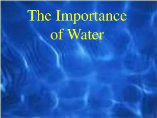
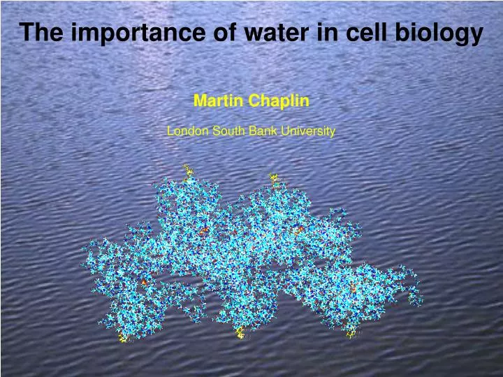
The importance of water in cell biology. Martin Chaplin. London South Bank University. The importance of water in cell biology. Outline of talk. Hydrogen bonding versus non-bonded interactions (conflict between enthalpy and entropy) Protein hydration

E N D
The importance of water in cell biology MartinChaplin London South Bank University
The importance of water in cell biology Outline of talk • Hydrogen bonding versus non-bonded interactions (conflict between enthalpy and entropy) • Protein hydration • Carboxylic acid clusters and the cytoskeleton • Intracellular water
Water: Structure Hydrogen atoms are not fixed Highly variable dipole moment and dielectric constant
Water: Structure No distinct lone pairs of electrons Hydrogen atoms are not fixed + - Highly variable dipole moment and dielectric constant Compact shape
Water: Structure No distinct lone pairs of electrons Hydrogen atoms are not fixed + - Highly variable dipole moment and dielectric constant Compact shape + Compact tetrahedral hydrogen bonding + - -
Water: Structure No distinct lone pairs of electrons Hydrogen atoms are not fixed + - Highly variable dipole moment and dielectric constant Compact tetrahedral hydrogen bonding Larger and non-spherical van der Waals shape
What is water’s hydrogen bond? Average values. In reality, there is much vibration and variation cf. van der Waals minimum energy position 3.0 - 3.6 Å
Water: Equilibrium Structure H-bond ~23 kJ/mol; O-H covalent bond ~492 kJ/mol O-H 0.97 Å O-H···O 1.88 Å Commonly found tetrahedral arrangement of water molecules. Hydrogen bonds O-H····O are not necessarily straight. Forms networks due to the substantial cooperativity in bond strengthening due to electron overlap within molecular orbitals.
Water: Equilibrium Structure Ubiquitous water tetrahedra
collapsed structure expanded structure Water: Equilibrium Structure
Water: Equilibrium Structure The most successful explanation for the special properties of water are found in the ‘Mixture models’ Dense clusters of water Lower density clusters of water
collapsed structure expanded structure Water: Equilibrium Structure maximizing van der Waals interactions more reactive maximising hydrogen bonds lower density more viscous
collapsed structure expanded structure Water: Equilibrium Structure maximizing van der Waals interactions more reactive maximising hydrogen bonds more viscous DH is -ve stronger bonds DS is -ve more ordered DG is ~0 finely balanced DV is +ve lower density Conflict between non-bonded interactions and hydrogen bonding Different conditions and/or solutes shifts the equilibrium
Water: Equilibrium Structure collapsed structure expanded structure a b Broad and shallow minimum Higher enthalpy but greater entropy Deep but localised minimum More negative enthalpy but smaller entropy Stronger hydrogen bonding; a b | Weaker hydrogen bonding; b a
Icosahedral dynamic equilibria Small clusters form larger clusters at lower temperatures. M. F. Chaplin, A proposal for the structuring of water, Biophys. Chem.83 (2000) 211-221.
Water: Equilibrium Structure DH is +ve DS is +ve DV is -ve expanded structure lower density water lower diffusion more viscous collapsed structure higher density water greater partner switching more reactive
Water: Equilibrium shifts expanded structure lower density more viscous collapsed structure higher density more reactive Cs+ > Rb+ > K+ Ca2+ > Li+ > Na+ ClO4- > H2PO4- > Cl- SO42-, HPO42- Leu, Ileu, Lys, Arg Asp, Glu Stabilise central dodecahedron Weakly hydrated ions with diffuse charge density. Stronglyhydratedwith high charge density. Ionic kosmotropes Ionic chaotropes
Protein hydration • Every amino acid hydrates differently: • Hydrophobic amino acids, e.g. leucine: low-density clathrate water surrounds • Basic amino acids, e.g. lysine: low-density clathrate water surrounds, some puckering • Hydrophilic amino acid, e.g. threonine similar to bulk water • Acidic amino acids, e.g. aspartic acid high density water surrounds with broken hydrogen bonding
Protein hydration Larger volume than average, lower density water Smaller volume than average, higher density water H. Zhao, Biophys. Chem. 122 (2006) 157–183.
Protein hydration Conflicting effects; mixed environments around proteins. Weak H-bonding allows greater flexibility. Strong H-bonding gives greater stability and solubility. Ordered structure in first shell around the protein, both hydrophobic clathrate-like and H-bonded; each helps the other to optimise water’s H-bonding network. Clathrate formation over hydrophobic areas maximises non-bonded interactions without loss of H-bonds. Carboxylate groups usually only fit a collapsed water structure creating a reactive fluid zone. Diffusion of surface water is only 10% of bulk water, and similar to supercooled water Protein rotation creates a surrounding zone of broken hydrogen bonds.
Rotational and translational diffusion Translational diffusion involves breaking water-water links at a distance from surface Rotational diffusion involves breaking close water-water and protein-water links. Interfacial region around a protein (perturbed water) comparable to protein volume The surface area for translation and rotation is the same but the velocity differential is constant for all r for translational but varies with r2 with rotation. At the breaking surface, half the H-bonds are broken. More hydration slows down rotation far more than effect on translation
Anchored proteins Static anchoring creates static surface water Static anchoring required to exert forces
The Cytoskeleton Actin, tubulin and intermediate filaments form the cytoskeleton in eukaryotic cells. Together they control mitosis, the shape of the cells and organise the cytoplasm and nucleus. Tubulin forms fat hollow and stiff microtubules making tracks for the movement of organelles. Actin forms thin flexible microfilaments. Intermediate filaments form flexible and elastic links. Actins all have acidic N-termini, Tubulins have acidic C-termini and Intermediate filaments have acidic central regions. The surface area of these filament systems exceeds that of all internal membranes 10-fold or more. They are highly conserved but their 3-D structures are known only in part. Fibres are known through electron crystallography and guesswork.
Actin Acidic N-terminus ADP/ATP binding site Actins are highly conserved with ~375 Amino acids. They form ~10% of total intracellular protein 1HLU Bovine b-actin profilin complex
Actin Filament Acidic N-terminus Actin filaments in the cytoskeleton are highly dynamic. Hydrolysis of ATP accompanies polymerization of ATP-containing monomers but destabilises the actin filaments. Water molecules shield the binding surfaces F-actin from Holmes KC and Eschenburg S
Actin N-terminus All known structures from UniProt Knowledgebase The a-actins have four terminal acidic groups whereas the b- and g-actins have three. The acid groups are conserved as either aspartate or glutamate. The amino terminal residue in many eukaryotic proteins is N-acetylated.
Selected Actins From mammals, birds, amphibia, crustacea and fungus a b / g a b / g a b / g a b / g
Tubulin b a b a GDP GTP GDP GTP + acidic C-terminus Tubulin has two similar subunits of ~450 amino acids. It has a GTP binding domain near the N-terminal, a beta-sheet core and alpha helices. Two antiparallel helices lead to the highly acidic external C-termini. There is a head to tail arrangement of dimers with the beta-subunit GTP at the open end. Only this GTP is hydrolysed following polymerization. 1Z2B Bovine a/b tubulin colchicine-vinblastine complex, introduces a curve not seen in native tubulin
Tubulin microtubules GDP GTP GDP GTP b a b a ~25 nm a b Acidic C-terminus Different numbers of subunits may coil round. The acidic negatively charged C-termini project into the external solution.
TubulinC-terminus All known structures from the UniProt Knowledgebase. The b-tubulins are mostly on the left.
Selected Tubulins From mammals, insects, plants, alga, fungi and protozoa a b a b a b a b
Intermediate filaments Fibrous elastic proteins formed mainly from coiled coils (‘ropes’) of multi-stranded acidic a-helices, ~11 nm diameter. The central ‘rod’ domains contain ~310 amino acids. Some acidic groups form salt links across to other strands but excess acidic groups are present on the surface of the ‘rod-like’ structure with excess basic groups at the end. Between a-helices are glutamate rich acidic bulges (single p-helices) forming flexible ‘linker’ regions. As strands gather these group together.
From mammals, birds, amphibia, snakes and fish, from cytokeratins, vimentins, desmins and neurofilaments Intermediate filaments Acidic p-loops
Acid oligopeptide clusters Acetyl-GLU-GLU-ASP- Acid groups tend to cluster together to share cations and to minimise water disruption
O C O Carboxylate effects +1 0 -1 -0.745 -0.774 2.23 Å chaotrope pKa=2.86 kosmotrope pKa=4.74 Causes density increase, if hydrogen bonded cf. sulfate (kosmotrope) –0.87, perchlorate (chaotrope) –0.71 Aspartic and glutamic acids are usually kosmotropic Different carboxylates have different pKas Na+ RCO2- pairs are always solvent separated (Na+ holds on to water strongly) K+ RCO2- pairs move from solvent separated to ion pair as pKa reduces 6-31G** basis set used
Carboxylate effects +1 0 -1 -0.745 -0.774 How can the pKa be shifted? chaotrope pKa=2.86 kosmotrope pKa=4.74
Carboxylate dipoles and pKa’s of the acids 2,2’ dimethylpropanoate 1.9 1.8 1.7 Carboxylate group dipole, D 1.6 1.5 trifluoroacetate 1.4 0 1 2 3 4 5 6 pKa Dipoles were calculated using ab initio molecular dynamics with the 6-31G** basis set, pKas from Dean, Lange's Handbook of Chemistry(1999).
Carboxylate effects +1 0 -1 -0.745 -0.774 How can the pKa be shifted? chaotrope pKa=2.86 kosmotrope pKa=4.74 By small changes in charge H-bonding to carboxylate increases O negative charge and pKa Clathrate structuring around carboxylate reduces O negative charge and pKa Overlapping negative field from nearby groups enhances counter ion association Ion pair association discourages hydrogen bonding but encourages clathrate formation 6-31G** basis set
Carboxylate effects High pKa, H-bonding to water Low pKa clathrate Ions dissociate on rotation Low density water High density water Static, fewer H2O collisions pulling it apart Ion binding Destroys signal Signals effect Solvent separated Na+, increasing charge Clathrate occupied ion pair K+, reducing charge
- - - - + + + + Na+ partitions away from low density water K+ prefers low density water Na+ does not shows magic number Na+(H2O)20 K+ shows magic number K+(H2O)20 Na+/K+ comparison Attraction Na+ - water > water – water > K+ - water + + Selective accumulation of K+ over Na+ where there is low density water Data from Collins KD, Biophys. J.72 (1997) 65, Millero FJ In Water and aqueous systems (1972), and Khan A Chem. Phys. Lett. 388 (2004) 342
Phosphates Intracellular concentration ~100 mequiv l-1 H2PO4- HPO42- pKa 7.21 6.67 Chaotrope zero ionic strength Kosmotrope >100 mM ionic strength As the ionic strength reduces the chaotropic H2PO4- concentration increases.
Transformation of water structuring ATP Free Protein Polymerized Protein ADP+Pi Static Greater H-Bonding = Low density water Rotation High density water = Broken H-bonds e.g.Rotating free G-actin F-actin filament If there is more order in the protein fibre, then there is more order in water Protein fibres trap water, which has decreased entropy. In order to attempt to keep the water activity constant, therefore, the water has to form bonds with a more negative enthalpy. This results in stronger bonds, causing greater structuring and lower density. Enclosure of water involving capillary action. This forms ‘stretched’ confined water is much more highly structured than the bulk water.
Transformation of water structuring water/water