Download
1 / 1
10 likes | 132 Views
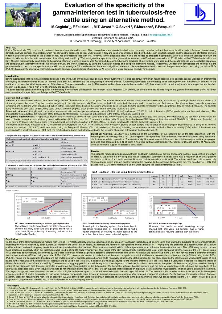
FIG.1 Data obtained according to different type of production. FIG.2 Data obtained according to the housing system. FIG.3 Data obtained according to age.

E N D
FIG.1 Data obtained according to different type of production FIG.2 Data obtained according to the housing system FIG.3 Data obtained according to age The obtained results according to the different categories showed that dairy cattle and dual purpose breed had a three times higher probability of resulting positive to the tests than beef cattle. According to the housing system, the animals kept in free-range housing and in mixed conditions had a higher probability of resulting M. bovis positive to the tests than the animals reared in tie stall system. Moreover, the results obtained according to age showed that 2-3 years old animals had a higher estimated risk of resulting positive than the others. M.Cagiola1*, F.Feliziani 1, M.T. Javed 3, G.Severi 1, P.Mazzone1, P.Pasquali 2 1*Istituto Zooprofilattico Sperimentale dell’Umbria e delle Marche, Perugia. e-mail: m.cagiola@pg.izs.it 2 Istituto Superiore di Sanità, Roma. 3 Department of Pathology, University of Agriculture, Faisalabad, Pakistan ABSTRACT Bovine Tuberculosis (TB) is a chronic bacterial disease of animals and humans. The disease has a world-wide distribution and in many countries bovine tuberculosis is still a major infectious disease among domestic and wild animals. The strategy, which has allowed the disease to be kept under control in Italy and in other countries, is based on the tuberculin skin test, isolation and the slaughtering of infected animals. In recent years the prevalence of the disease in Italy has decreased to less than 1%. However, further diagnostic tests in vivo are needed to eradicate it completely. Such tests could be used together with the tuberculin skin test in order to increase levels of sensitivity and specificity. We investigated the specificity of the gamma-interferon test in 800 animals selected from 20 officially certified TB-free herds in Umbria, Italy. The skin test specificity was 96.8%. In the gamma interferon testing, in parallel with Australian tuberculins, tuberculins produced at our Institute were used and the results obtained were evaluated separately and comparatively (alternative method). We obtained 97.3% and 98.6% specificity by using the Australian method and using the alternative method, respectively. Our research corroborated the findings that the gamma-interferon test is more specific than the skin test to investigate TB prevalence and there was a significant statistical difference between the skin test and the gamma-interferon test using the Italian PPDs (P=0.03). Moreover the alternative method allowed us to discover a higher degree of positivity for M. avium and a lower degree of positivity for M. bovis Evaluation of the specificity of the gamma-interferon test in tuberculosis-free cattle using an alternative method. Introduction Bovine tuberculosis (TB) is still a widespread disease in the world. Not only is it a serious obstacle for productivity but it is also dangerous for human health because of its zoonotic aspect. Eradication programmes are ongoing in several countries based on the use of the skin test, isolation and the slaughtering of infected animals. Further diagnostic tests are necessary to be used together with the tuberculin skin test for the eradication in countries with low prevalence of the disease. The gamma-interferon test (-IFN) as other authors have already demonstrated (6,7), has turned out to be extremely useful as a supportive test in vita to the skin test because it has a high level of sensitivity and specificity (3). The same test has been a determining factor in eliminating the outbreaks of infection in the Northern Italian Regions (1). In Umbria, an officially-certified TB-free Region, the gamma-interferon test (-IFN) has been used to evaluate its specificity in officially-certified TB-free herds. Materials and Methods Animals: 800 animals were selected from 20 officially-certified TB-free herds in the last 10 years.The animals were found to have paratuberculosis-free status, as confirmed by serological tests and the absence of clinical signs over the years. They had reacted negatively to the skin test and only 25 of them resulted dubious to both the single and comparative test. Furthermore, the aforementioned animals showed no symptoms and no lesions when slaughtered. When further tests were carried out on the organs which had been removed from the animals immediately after slaughtering, they all resulted negative. The animals being tested were beef cattle (n°550), dairy cattle (n°100) and dual purpose breed (n°150) with different housing systems and ages. Tuberculin skin test: A single and comparative intradermal skin test was performed using bovine (50.000 I.U. /ml) and avian (25.000 I.U./ml) tuberculins (PPDS) produced at our licensed laboratory. The protocols for the production, the execution of the test and its interpretation were carried out according to the criteria previously reported (8). The gamma interferon test: A heparinised blood sample (10 ml) was collected from each animal just before carrying out the tuberculin skin test. The samples were delivered to the lab within 8 hours from the blood collection, using the method already described by others (4,5). Each sample (1.5 ml ) was stimulated with: 30 g of Australian bovine PPD, 30 g of Australian avian PPD (CSL Ltd., Melbourne, Australia), 10 g of bovine PPD and with 10 g of avian PPD produced at our Institute. A solution of PBS (0.01M, pH 7.2) was employed to estimate the basal value of -IFN. The blood samples were incubated at 37 °C in a humidified atmosphere (5% CO2) for 16-24 hours. After incubation, plasma of each sample was collected by centrifuging the blood cultures at 500g for 10 minutes at room temperature (22° ± 5°C) and it was tested using the Bovigam ELISA test (CSL Ltd., Melbourne, Australia), following the instructions included in the kit. The optic density (O.D.) value of the results was measured with a spectrophotometer (450 nm).The results obtained were evaluated according to the following alternative criteria described by others (4,5): Statistical Analysis. Specificity was measured as the percentage of true negative out of the total population with the following formula: true negative x 100 / (true negative + false positive). The confidence limits were calculated using the normal distribution for infinite populations. The statistical significance of several differences, found in the analysis and the grouping of data was also calculated. EPI INFO 2000, a free-ware software distributed by the Center for Disease Control of Atlanta was used as electronic support for statistical calculations. I interpretative level: separate evaluation of data obtained after stimulation with Aust. and Ital. PPDs Results The separate results obtained using Australian and Italian tuberculins at the first and second levels of interpretation are shown in Table 1. We noted that by using also Italian tuberculins (alternative method) there was a reduction of M. bovis positive animals from 21 to 10 and an increase of M. avium positive animals from 44 to 54. The animals confirmed dubious were only 10 (non discriminative animals) and they all resulted negative to the succesive tests carried out. The specificity of -IFN test passed from 97.3% at the first level to 98.6% when applying the second interpretative level.. II interpretative level: comparison of separate data obtained after stimulation with Aust. and Ital. PPDs Tab.1 Results of -IFN test using two interpretative levels (Goria M. et al, 1997) Conclusion On the basis of the obtained results we noted a high level of -IFN test specificity with values between 97.3% using only Australian tuberculins and 98. 6 % using also tuberculins produced at our licensed Institute, exceeding the values reported by other authors (2). Moreover the use of Italian tuberculins reduced the number of false positive animals from 21 to 11, highlighting the presence of a higher number of M. avium positive animals, and confirming only 10 dubious animals (non discriminative reaction). The above data underlined that different parameters can influence the results of the tests. The -IFN assay tends to reduce these influences, especially when our tuberculins were used to stimulate blood lymphocytes. The values of the skin test specificity recorded were lower when compared with the values of the -IFN assay using PPDS produced at our Institute, as confirmed by the 2 test.There was neither a significant statistical difference between the use of Australian and Italian PPDs in the -IFN test nor was there a difference between the skin test and the -IFN test using Australian PPDs (P=0.07). However we wanted to underline that there was a significant statistical difference between the skin test and the -IFN test using Italian PPDs (P=0.03). Taking into consideration this data and the limited number of animals observed (which could negatively influence the statistical results), our study could be the starting point which might trigger off and lead to further research on the best choice of tuberculins to be used in the -IFN test. In addition, our findings showed for the first time that the use of the -IFN test is a useful tool to reduce the negative effects of different factors which can influence specificity. These results strongly suggest that a possible way to improve diagnostic procedures, in order to better control the spread of tuberculosis, might be based on the right choice of the diagnostic test, focusing particular attention on the characteristics of the tuberculins. We found that different housing systems and the type of production could influence the specificity of the tuberculosis diagnostic tests. Even though our results do not shed light on the reason for this, we can suppose that it depends on exposure to environmental mycobacteria, which is able to sensitize the animals. With regard to age, we noted that the risk of sensitization is higher in the cows aged 2-3 and 4-5 years old than in the cows aged 6-7 years old. The reason for this, as other authors have reported, is the complex antigenic structure of bovine tuberculin, in that some particular components could induce an immune-suppressive response after repeated intradermal injections. On the basis of the results obtained from our research, especially regarding the high level of specificity (98.6%) derived from using the tuberculin produced at our Institute in the gamma-interferon test, we can conclude that the -IFN assay would be a valid diagnostic aid, together with the skin test, in the final phases of eradication programmes. References 1. Archetti I.L., Amadori M., Scaccaglia P., Vezzoli F., Luini M., Fai M., Belloli A., Sala L.(1996). Impiego del test -interferon per la diagnosi di tubercolosi bovina in regione Lombardia. La Selezione Veterinaria 4,329-338. 2. Dondo A. & Goria M. (1998). Esperienze nell’impiego della prova del gamma interferone. Medicina Veterinaria Preventiva (supplemento 1998),41-46. 3. Dondo A., Goria M., Moda G., Cesano L., Garanzini A., Giammartino M., Minola G., Moriconi E., Porta G., Banchio P., Marmo G. (1996). La prova del -interferone per la diagnosi della tubercolosi bovina: determinazione della sensibilità e della specificità in prove di campo. Medicina Veterinaria Preventiva, 13,14-18. 4. Dondo A. & Goria M.(1997). Diagnosi in vita della tubercolosi bovina mediante -interferon test. “Infezioni da micobatteri tubercolari e non tubercolari negli animali e nell’uomo: attualità e prospettive future”. Atti del Congresso.74-82. 5. Goria M., Faccenda L., Bianco C., Gemello P., Pizzoni E., and Dondo A.(1997). Impiego del - IFN test nella diagnosi di tubercolosi bovina in allevamenti infetti da M. paratuberculosis. Il Progresso Veterinario.11/97: 399-401. 6. Lauzi S., Pasotto D., Amadori M., Archetti I.L., Poli G., Bonizzi L. (2000). Evalutation of the specificity of the -interferon test in Italian bovine tuberculosis-free herds. The Veterinary Journal 160,17-24. 7. Wood P.R. & Rothel J.(1994). In vitro immunodiagnostic assays for bovine tuberculosis. Vet. Microbiol. 40 (1-2):125 –35 8. Anonymous (2000). Manual of standards for diagnostic tests and vaccines of World Organization for Animal Health (OIE), 4th edition