Download
1 / 22
230 likes | 521 Views
Arterial system Pressures in the circulation Arterial pressure during diastole Pulse pressure and work of the heart Hemodynamics is the study of the physical principles that govern blood flow in the cardiovascular system. Factors affecting resistance to flow Laminar & turbulent flow

E N D
Arterial system • Pressures in the circulation • Arterial pressure during diastole • Pulse pressure and work of the heart • Hemodynamics is the study of the physical principles that govern blood flow in the cardiovascular system. • Factors affecting resistance to flow • Laminar & turbulent flow • Tension, pressure and radius Arterial System & Hemodynamics
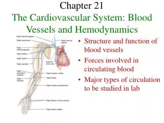
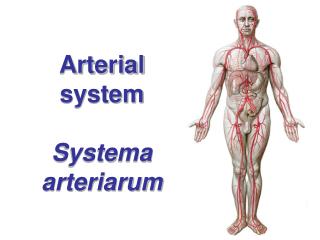
Arterial system The arterial system consists of: Elastic arteries: major distribution vessels with a large component of elastic tissue and low resistance: aorta, brachiocephalic, common carotid, subclavian and pulmonary arteries. Muscular arteries: distributing branches with more muscular tissue and less elastic tissue: radial, femoral, coronary and cerebral arteries. Arterioles: terminal branches that supply the capillaries.
= MAP Systemic Pulmonary Arteries L ventricle 120 Rt ventricle Capillaries Capillaries L ventricle Arterioles Rt atrium Pulse pressure Venules L atrium 100 Arteries Veins Veins 80 Pressure, mm Hg 60 Diastolic pressure 40 20 Shaded area = systolic pressure - diastolic = pulse pressure Systolic pressure Mean & pulse pressures in the circulation
Peak systolic pressure & pulse pressure are determined by stroke volume & aortic compliance. Compliance depends on the elastic tissue of the aorta. Pulse pressure depends on stroke volume and aortic compliance Aortic valve closure Stroke volume Pulse pressure Mean arterial pressure Aortic compliance Pulse pressure = peak systolic minus diastolic pressure. A decrease in aortic compliance will result in a higher peak systolic pressure & pulse pressure (assuming stroke volume is unchanged).
Aorta systole pressure Left Ventricle diastole The stroke volume is ejected during the rapid ejection period & is accommodated by expansion of the aorta. Aortic pressure is maintained during diastole by recoil of the aorta as blood flows to the periphery. Because of the elasticity of the aorta and large arteries, the pulsatile pressure signal is gradually dampened so that flow is steady (not pulsatile) in the venules. Arterial pressure is maintained during diastole by recoil of the aorta During diastole the aortic valve is closed and recoil of the elastic aorta drives blood to the periphery
Central and peripheral aortic pressures Simultaneously recorded pressures from the aortic root (Ao) and femoral artery (FA) demonstrate delayed transmission and a higher systolic pressure in the femoral artery. Although peak pressure is higher in the femoral artery than the aorta, average driving pressure (MAP) is higher in the aorta than the femoral artery. There is smoothing of the pressure waveform and loss of the dicrotic notch between the aorta and femoral artery. UpToDate®
Work = force (f) operating over distance (dl): W = (f)( dl) For work done by a piston with area A moving against pressure P, For the left ventricle: P = afterload = arterial pressure when the aortic valve is open & V = stroke volume, so cardiac work = stroke volume x afterload A P dl The work of the heart consists of pumping volume against pressure
Determinants of pulse pressure: Stroke volume Aortic compliance Aging & atherosclerosis aortic compliance systolic pressure & pulse pressure cardiac work due to systolic pressure Systolic and pulse pressures increase with age systolic 150 mean Arterial pressure 100 diastolic 50 60 20 40 80 Age
C = DV/DP 250 200 22 years old % increase in volume 150 75 years old 100 50 60 100 140 180 Pressure, mm Hg Cardiac work = stroke volume x afterload. Cardiac work stroke volume x systolic BP. Increasing systolic pressure increases cardiac work. Compliance of the aorta minimizes peak systolic pressure & cardiac work. Cardiac work increases with age, & in hypertension, aortic stenosis or coarctation. A decrease in aortic compliance or an increase in afterload increase cardiac work Difference in aortic compliance in young versus old subjects
The resistance of an individual vessel is inversely proportional to radius so R artery < R arteriole < R capillary The total resistance of a category of vessels is determined by the total cross sectional area of all the vessels and the radius of the individual vessels. Comparing arteries & arterioles supplying an organ, the total resistance of the arterioles is greater than the resistance of the arteries. Comparing arterioles & capillaries, the total cross sectional area of the capillaries is so much greater than the area of the arterioles that the total resistance of the capillaries is less than the resistance of the arterioles. The greatest pressure drop in the circulation is across the arterioles. Arterial, arteriolar & capillary resistance
Conductance is the inverse of resistance. For Parallel circuits conductances are additive Ctotal = C1 + C2 + C3 Series and parallel resistance Series & parallel resistances Resistances in series are additive; total resistance equals the sum of individual resistances. Resistances in parallel add as the inverse sum, like parallel electrical circuits: Most vessels of a given category are arranged in parallel.
Coronary arteries Arteries to CNS Arteries to limbs & trunk Aorta Arteries to stomach, spleen, pancreas, gut Hepatic artery Renal arteries Arterial beds are arranged in parallel Venous pressure is small compared to arterial pressure. Blood pressure is nearly the same in all large arteries, so DP is the same in all vascular beds: DP = MAP – VP MAP and MAP = CO X TPR The resistance of each arterial bed is set by its own arteriolar tone TPR is less than the resistance of any single arterial bed. For example: TPR = MAP/CO Renal resistance = MAP/renal flow And renal flow < CO so Renal resistance > TPR There are multiple parallel paths for blood flow so total peripheral resistance is less than resistance in any one bed. Parallel resistances in the systemic circulation
Normal flow in the circulation is laminar. Laminar (or streamlined) flow exhibits maximal velocity at the center of the vessel, and concentric thin layers of plasma with gradually decreasing velocity toward the walls of the vessel. Laminar flow is silent. Plasma flowing closest to the vessel wall exerts a drag on the wall (shear stress) that influences endothelial function. Laminar & turbulent flow Laminar Turbulent flow exhibits irregular radial mixing of blood as overall flow occurs in the longitudinal direction. Turbulent flow usually causes vibrations that are audible with the stethoscope as murmurs or bruits. The pressure gradient required to drive turbulent flow is greater than that required to drive laminar flow so turbulence increases cardiac work. Turbulent
Flow in the vascular system is normally laminar. Turbulence occurs if Reynold’s number (NR) exceeds 3000: Factors predisposing to development of turbulence in flowing fluid include: Greater density (r) larger vessel diameter (D) High velocity (v) Low viscosity (h) In addition, turbulence is likely in the presence of abrupt changes in vessel diameter or irregularities in vessel walls as may occur with atherosclerosis or other pathology. Causes of turbulent flow Examples of turbulent flow: Flow across an obstruction (aortic stenosis, coarctation) Abnormally high flow velocity (high CO & reduced viscosity in severe anemia) Regurgitant flow across an incompetent heart valve Abnormal shunt from a high to low pressure chamber (ventricular septal defect) Presence of turbulent flow increases the likelihood of development of blood clots
Normal hematocrit Resistance to blood flow is proportional to viscosity (): 10 30 50 70 Viscosity of a fluid is its resistance to flow resulting from molecular cohesion. Blood is a complex mixture of fluid and cells; blood viscosity increases with hematocrit. Viscosity and hematocrit Viscosity of whole blood (solid line) relative to plasma (dashed line) as a function of hematocrit polycythemia viscosity anemia Hematocrit
0.1 0.3 0.2 0.4 0.5 Blood viscosity decreases in vessels with diameter < 0.3 mm (300 mm) Mechanism is complicated but the effect reduces resistance to blood flow. Most of the resistance in the systemic circulation is from arterioles with small diameters. Lower resistance reduces the work of the heart. Viscosity increases at low temperature, & may reduce blood flow in the extremities & contribute to frost bite. Vessel diameter and viscosity of blood Relative viscosity Vessel diameter, mm
The law of Laplace states that tension (T) in the wall of a blood vessel equals the product of transmural pressure (P) and radius (r): Tension is a force acting tangential to the surface of a cylinder. P is transmural pressure (internal pressure minus external) Transmural pressure equals blood pressure minus tissue pressure. Extravascular tissue pressure is small & can be ignored so P T Law of Laplace
Law of Laplace and tension in blood vessel walls T = Pr The amount of elastic tissue in the vessel wall correlates with the wall tension. Elastic tissue maintains wall integrity against the outward force of the pressure. The table shows that a very small tension in the wall of the capillaries suffices to withstand the intraluminal pressure. For comparison, the breaking strength of a strip of Kleenex one cm wide is about 50,000 dynes per cm, over 3000 times as great as the tension required to maintain a capillary wall.
Slope = Compliance = DV/DP 400 300 Relative volume, % 200 Compliance decreases slightly at higher pressures 100 50 100 150 200 Pressure, mm Hg Compliance of the aorta as an example of an elastic artery In this figure smooth muscle is relaxed pharmacologically so the vessel’s compliance is due primarily to the compliance of the elastic tissue. Compliance of elastic arteries is nearly constant over the physiological range of pressure.
Slope = Compliance = DV/DP 400 300 Relative volume, % 200 Compliance decreases as cross section becomes circular 100 50 100 150 Pressure, mm Hg Maximal physiological pressure Compliance in the vena cava as an example of a large vein In this figure smooth muscle is relaxed; the compliance is due primarily to the changes vessel geometry.
100 120 140 160 180 Collagen fibers only Contribution of collagen & elastic fibers to passive tension development T= Pr 160 Intact artery 120 Collagen fibers are least compliant, develop greatest tension for a given radius. Tension, dynes/cm x 103 80 elastic fibers only 40 Relative radius, % Passive tension development (smooth muscle relaxed) with increasing radius in an intact artery or an artery with only collagen fibers (elastic fibers digested) or only elastic fibers (collagen fibers digested). Arterial segment studied in vitro by injecting fluid & measuring radius & tension.
T = Pr Arteries: If weakening of the wall causes dilatation (aneurism) , as the radius increases the tension required to maintain wall integrity increases. If the wall is too weak to maintain the tension, it will rupture. The heart: Pathological dilation of the heart as occurs in heart failure increases the radius of the ventricles. As a result, more tension must be generated to create a given pressure, increasing the work of the heart & causing progression of heart failure. Wall tension and pathological changes