Download
1 / 55
600 likes | 1.05k Views
Normal ECG: Rate and Rhythm. Read chapters 4 and 22. ECG Interpretation*. Standardization Rate RR interval Heart rate Rhythm PP interval P wave width, height, shape, etc. PR interval QRS width (and height) axis R wave progression abnormal Q waves ST segment T waves

E N D
Normal ECG: Rate and Rhythm • Read chapters 4 and 22
ECG Interpretation* • Standardization • Rate • RR interval • Heart rate • Rhythm • PP interval • P wave • width, height, shape, etc. • PR interval • QRS • width (and height) • axis • R wave progression • abnormal Q waves • ST segment • T waves • QT interval • U waves *See Chapter 22
ECG Interpretation • Univ. of Wisconsin Medical School • http://www.fammed.wisc.edu/pcc/ecg/
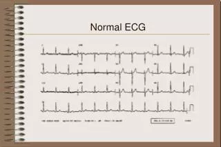
The Normal ECG • Normal = normal sinus rhythm
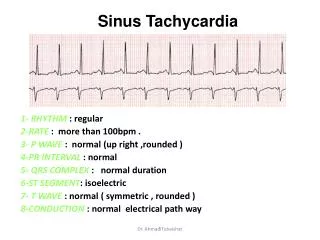
Rate • R-R interval • Is it regular? • What is the heart rate? • 300, 150, 100, 75, 60, 50 • 300 / (# of large boxes) • 1500 / (# of small boxes) • Count the number of cardiac cycles in 10 seconds and multiple by 6.
Rate • Bradycardia • less than 60 bpm • Tachycardia • greater than 100 bpm
Rate • P-P interval
Rhythm • P wave • PR interval • QRS
4. P Wave • Lead II and aVR • Positive in II • Negative in aVR • < 2.5 mm in amplitude • < 0.12 sec. in width
Normal P Wave Normal direction of atrial depolarization aVR? II? Figures 4-2 and 4-3
Abnormal P Wave Direction of atrial depolarization with junction rhythm aVR? II? This is an example of a retrograde conduction
P wave • The same direction as QRS • Only one P wave in front of QRS • Do all the P waves look alike?
5. PR interval • 0.12 - 0.20 seconds
6. QRS Complex • What is the width? (less than 0.10 seconds) • Do all the QRS waves in the same lead look alike? • R wave progression • Axis • Abnormal Q waves (infarction)
QRS Complex Q waves
Normal QRS • Two phases • brief phase; depolarization of ventricular septum • longer phase; depolarization of both ventricles but the left is larger
First Phase • Depolarization of ventricular septum
Second Phase • Depolarization of both ventricles but the left is larger
Precordial Leads V6 V1
Normal QRS V6? V6? V1? V1? Fig. 4-6
Normal QRS V1 V6
Normal QRS • Septal r wave • Septal q wave
6. QRS Complex • R wave progression
Normal R Wave Progression Transition Zone?
R Wave Progression Transition Zone?
Transition Zone • Figure 4-7
Early & Delayed Transition V1 V2 V3 V4 V5 V6 • Figure 4-7
6. QRS Complex • What is the electrical axis? • normal • left axis deviation • right axis deviation • extreme axis deviation
7. St Segment • ST segment elevation or depression • (see chapters 8 & 9)
8. T Wave • Normally positive where QRS wave is positive • V3- V6 and II,but negative in aVR • Abnormally tall T waves
Practice • ECG Library • http://www.ecglibrary.com/ecghome.html • ECG: The Art of Interpretation • http://www.12leadecg.com/full/
Not normal PR interval - Mobitz Type II block
Not normal LAD, R wave progression RBB w/inferior MI
Not normal - First degree block, left atrial enlargement, left bundle branch block, & inferior MI
Not normal Atrial fibrillation
Not normal Junctional rhythm
Not normal LAD, late R wave progression Acute MI
Not normal Premature ventricular contractions
Not normal Ventricular tachycardia: note fast rate and wide bizarre QRS.
Not normal Second degree AV block - type II
Not normal RAD, R wave progression
Not normal Third degree AV block