Download
1 / 13
130 likes | 408 Views
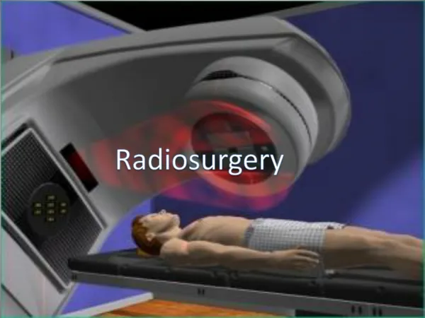
Optimizing Gamma Knife Radiosurgery through Mathematical Morphology. Jeffrey Overbey Angela Kolve Nathan Hirtz. What is Gamma Knife Radiosurgery?. Used to treat brain tumors. Delivers a high dose of ionizing radiation from 201 cobalt-60 beams.

E N D
Optimizing Gamma Knife Radiosurgery through Mathematical Morphology Jeffrey Overbey Angela Kolve Nathan Hirtz
What is Gamma Knife Radiosurgery? • Used to treat brain tumors. • Delivers a high dose of ionizing radiation from 201 cobalt-60 beams. • These beams intersect in an approximately spherical shape. • Each dose of radiation is called a shot.
What is Gamma Knife Radiosurgery? • Beams emanate from collimator helmet. • 4 interchangeable outer helmets. • Beam sizes are 4, 8, 14, or 18mm in diameter. • The target point, or center of the shot, is called the isocenter. http://www.mc.uky.edu/gammaknife/images/gdraw.jpg
What is Gamma Knife Radiosurgery? • The “edge” of each shot is called the 50% isodose line (50% IDL). • Beyond the 50% IDL, the radiation level is less than 50% of that at the isocenter, and is not considered damaging. http://w3.uokhsc.edu/neurosurgery/gamma/gamkni2.jpg
What is Gamma Knife Radiosurgery? http://www.ucsf.edu/gammakf/lgk_cutout2.jpg http://www.erheadquarters.com/episodes/8/images/allinhead2.jpg
The Problem • Given a volume, find the most optimal method for arranging spheres of 4, 8, 14, and 18mm in diameter given the following conditions: • At least 90% of the volume is occupied. • No spheres overlap. • No spheres protrude outside the target volume.
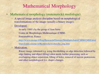
Mathematical Morphologyand Skeletonization • Morphology is a field of study with applications in computer vision, handwriting recognition, and image processing. • Skeletonization is a morphological process that reduces an image to its most basic linear structure.
Skeletonization http://www.cee.hw.ac.uk/hipr/html/skeleton.html
Skeletonization http://www.esiee.fr/~coupriem/Sdi/resources/saha3_SC.gif
The Formula and Its Graph 0.7717657102 – 0.04046790807x + 0.2693164541y z = 1 – 0.1580097209 ln x – 9.032235572 ln y x = number of shotsy = percentage treatedz = preference value
Analysis of the Model • Limitations • The shapes of the shots are not perfectly spherical. • The formula is based on a certain set of preferences.
Analysis of the Model • Benefits • At least 90% of the target volume is covered. • Largely confines untreated tissue to a small area. • Overlapping areas of strong radiation from 2 or more shots is avoided. • Represents a compromise between minimum number of shots and area of target volume covered.