Download
1 / 34
350 likes | 705 Views
HEMATOLOGIC SYSTEM. Ch. 14 Goodman. OVERVIEW. *The hematologic system involves the blood, blood vessels and the associated organs. *The study of hematology includes the diseases of the hematological system.

E N D
HEMATOLOGIC SYSTEM Ch. 14 Goodman
OVERVIEW • *The hematologic system involves the blood, blood vessels and the associated organs. • *The study of hematology includes the diseases of the hematological system. • * This critical system is tightly integrated with the following systems: lymphatic, immune, circulatory, respiratory. It is also integrated with hormonal and metabolic functions. • ***While there are primary diseases of the hematological system, the most common conditions are most often signs of other conditions
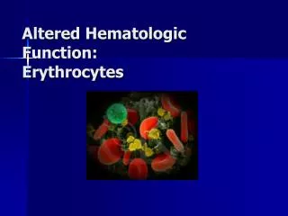
COMPONENTS OF BLOOD • *2 components of blood: • Plasma • Formed elements • Erythrocytes (RBCs) • Leukocytes (WBCs) • Thrombocytes (platelets)
SIGNS OF HEMATOLOGICALDISORDERS • Edema (sign) Excessive fluid accumulation within the interstitial tissues or within body cavities • Infarction (sign) localized region of necrosis caused by reduction of arterial perfusion • Thrombus (sign) blood clot - solid mass within intact vessel or the heart • Embolus (sign) Thrombus on the move that gets lodged distant from its place of origin
MORE SIGNS…. • Lymphedema (sign) Can be a sign of hematologic conditions - increased lymphatic load associated with decreased plasma proteins • Bleeding / bruising (sign) A sign when it happens with minor trauma or if bleeding continues longer than normal • Shock (sign) Inadequate blood pressure to perfuse organs
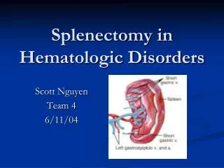
AND STILL MORE… • Lymphedenopathy (sign) Abnormal enlargement of lymph nodes • Splenomegaly enlarged spleen (spleen removes old RBC’s and antibody-laden bacteria or cells)
AGING/HEMATOPOIETIC SYSTEM • Aging and the Hematopoietic System • Decreased red marrow • Decreased intestinal iron absorption • Increased fragility of plasma membranes • Increased fibrinogen and platelet adhesiveness • Earlier activation of coagulation system • (ergo…) disturbed blood flow, propensity for anemia (low RBCs) - if nothing else slower RBC recovery from a loss of blood
BLOOD TRANSFUSION REACTIONS • Blood Transfusions • Reaction to blood and blood products • 1. Febrile nonhemolytic reaction - most common (.5-1% of transfusions), fever (at least 1 degree rise) but stable cells • 2. Transfusion related acute lung injury - 1 in every 2000 transfusions; initially mild shortness of breath but can progress to appear clinically as acute respiratory distress syndrome (widespread inflammation, low ventilatory volumes, poor oxygenation); with treatment can prevent / minimize permanent lung damage • 3. Acute hemolytic transfusion - 1 in every 25,000 transfusions - severe response due to ABO incompatibility; RBCs are destroyed (lysis) - mortality rate is high (17-60%)
BLOOD TRANSFUSION REACTIONS (CONTINUED): • 4. Allergic reactions - 1-3% of all transfusions, most common with fresh frozen plasma and platelet transfusions - allergic reaction • 5. Anaphylaxis - 1 in 20,000 transfusions - acute onset of hypotension, laryngeal edema • 6. Sepsis - very rare; due to bacterial contamination of blood products used in transfusion • Bloodless medicine - goal to reduce blood loss with procedures to avoid need of transfusions
DISORDERS of IRON ABSORPTION • Disorders of Iron Absorption • Hereditary Hemochromatosis is an autosomal recessive hereditary disorder characterized by excessive iron absorption by small intestine • Uncoupling of absorption and needs - leads to iron deposition in cells, particularly the liver, pancreas and heart • Early signs include weakness, hepatomegaly, elevated liver enzymes; symptoms include myalgias, joint pain, fatigue
Hereditary Hemochromatosis (continued) • Diagnosis-by blood work • Treatment - therapeutic phlebotomy • Prognosis -is good (from a mortality / morbidity perspective) but the condition is not reversible - it is managed
DISORDERS OF ERYTHROCYTES • Anemia • 1. Definition • reduction in oxygen carrying capacity of blood due to reduced quantity or quality of RBCs • HgB < 14 g/100 ml for men; 12 g/ 100 ml for women
Anemia (continued) • 2. Overview • Not technically a disease - really a sign of other underlying disorders - including but not limited to: • ** dietary (folate, vitamin B12) • **acute or chronic blood loss • ** iron deficiency (diet or absorption) • ** congenital defects (sickle cell anemia) • **poison exposure • ** disease of bone marrow • **chronic inflammatory, infectious or neoplastic disease • ** any disorder that upsets the balance between blood loss through bleeding or destruction and production of RBCs
Anemia (continued) • 3. Clinical manifestations • Mild - minimal and vague symptoms of fatigue • Moderate to severe progression: weakness, dyspnea on exertion, tachycardia, increased angina in pre existing CAD, dementia • 4. Treatment • a. Underlying problem (cause) if possible • b. Blood transfusions
DISORDERS OF LEUKOCYTES • Disorders of Leukocytes A. Leukocytosis ** Definition & Etiology • Increased number of leukocytes (WBCs) for a variety of causes - including as a normal response stressors • Common finding with infection - > 10,000 WBCs/mm^3 **Clinical manifestations • Signs and symptoms are usually those of infectious conditions (localized or systemic) - fever, headache, shortness of breath ** Treatment Underlying problem
Disorders of Leukocytes • B. Leukopenia **Definition & Etiology Decreased number of WBCs - < 5000 / ml caused by a variety of conditions: * HIV, hepatitis *alcohol * Nutritional deficiencies *connective tissue disorders (SLE) * bone marrow failure (i.e. following antineoplastic chemotherapy) **Clinical manifestations Asymptomatic; increased risk of infection **Treatment Underlying problem
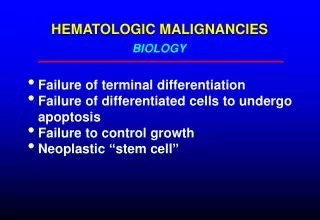
NEOPLASTIC DISEASES OF THE BLOOD AND LYMPH SYSTEMS • **** Bone marrow transplant Treatment of choice for any hematologic neoplastic disease • A. Leukemias • Malignant neoplasm of blood forming cells that replaces normal bone marrow with a malignant clone • Acute vs. Chronic • Myeloid vs. Lymphocytic • Blastic vs. cytic • Acute progresses quickly; chronic slowly • Myeloid - bone marrow origin involving hematopoeitic stem cells • Lympho - involving lymphoid or lymphatic system • Blastic - large, immature (functionless) cells • cytic - immature, smaller cells • Immunologically can also classify as T-cell / natural killer cell and B-cell leukemias
Leukemia (continued) • Three main clinical consequences: • 1. Anemia • 2. Infection • 3. Bleeding tendencies • Acute myelogenous leukemia - AML - most common leukemia in adults • Acute lymphoblastic leukemia - ALL – diagnosed most commonly in children • Chronic myeloid leukemia - CML – occurs most often in adults • Chronic lymphocytic leukemia - CLL – a common type of adult leukemia
MALIGNANT LYMPHOMAS • Malignant Lymphomas • Cancers of lymphatic system • Two groups - Hodgkin’s lymphoma and Non-Hodgkin’s Lymphoma (HL and NHL) - although the distinction has become less clear • More useful to categorize based on cell behavior - indolent vs. aggressive • HL is distinguished from others by presence of a cell known as the Reed-Sternberg cell in the lymph nodes
Hodgkin’s Lymphoma • HL - adults and children; B-cell malignancy; Clinical manifestations are variable, Box 14-6, Ann Arbor Staging Classification for HD (Hodgkin’s Disease) - progresses by increased involvement of lymph nodes and other lymph organs; Stage IV includes involvement of extralymphatic organs (liver, lung, skin)
Non-Hodgkin’s Lymphoma • NHL - large group (~30 types) of lymphoid malignancies that present as solid tumors arising from lymphatic cells - 5th most common type of cancer in the US with an incidence of ~67,000 / year; clinical manifestations are variable
MULTIPLE MYELOMA • Multiple Myeloma • MM - primary malignant neoplasm of plasma cells arising in bone marrow • Progression causes damage to kidney, recurrent infections, often affects the nervous system • Increased incidence (doubled in past 2 decades) (~16,000 new cases / year) - thought in part to increase in population > 85 years old
MYELOPROLIFERATIVE DISORDERS • Myeloproliferative Disorders • Originate from a hematopoietic stem cell that has transformed to allow the cell to mature and function with uncontrolled function - common characteristics include: • 1. hypercellular bone marrow • 2. tendency to thrombosis and hemorrhage • 3. increased risk of evolving into acute leukemia
Types of Myeloproliferative Disorders • Polycythemia vera - increased production of RBCs • Essential thrombocythemia - most common - increased platelet count
DISORDERS OF HEMOSTASIS • ***The following 2 processes occur normally in response to tissue insult and bleeding • Primary hemostasis - formation of a platelet plug • initiated when collagen fibrils and von Willebrand’s factor (vWF) in the subendothelial matrix of the blood vessel get exposed to blood • Secondary hemostasis–formation of fibrin through the coagulation cascade • vascular damage exposes ‘tissue factor’ which initiates the cascade
Disorders of hemostasis • A. von Willebrandʼs Disease • Most common inherited bleeding disorder - • Disorder of primary hemostasis - mucosal and skin bleeding and prolonged oozing after trauma or surgery • Rx - replacement of vWF
Disorders of hemostasis • B. Hemophilia • Deficiency of clotting factors • Disorder of secondary hemostasis - easy bruising, prolonged bleeding • If severe can spontaneously bleed - into joints, muscles, organs • Rx - replacement of clotting factors
Disorders of hemostasis • C. Thrombocytopenia • Decreased platelets (< 150,000/mm^3) - caused by either reduced production or increased destruction; usually a sign of other problems • Mucosal bleeding is a common sign • May be presenting sign of aplastic anemia (bone marrow failure) • Rx - underlying cause; platelet transfusions
Disorders of hemostasis • D. Aspirin / NSAIDs • Single dose of ASA can impair platelet function for 48 hours • NSAIDs are less potent • ASA and NSAIDs are contraindicated before surgery
Disorders of hemostasis • E. Disseminated Intravascular Coagulation • DIC - thrombotic disease caused by overactivation of the coagulation cascade - • Paradoxical in that clotting and hemorrhage occur simultaneously within the vascular system • Widespread clotting and fibrin deposition in circulation, causes backflow and increased pressure which leads to hemorrhage • Common after shock, sepsis, obsteteric / gynecological complications, cancer, massive trauma • Alteration in normal balance of pro and anticoagulant factors • Rx - underlying cause
Disorders of hemostasis • F. Hemoglobinopathies • Abnormalities in the formation of HgB • 1. Sickle cell disease (SCD) - • Genetic, result is HgB that changes from biconcave to crescent (sickle) shape once oxygen is released but leads to difficulty in blood flow in microvascularareas resulting in acute and chronic tissue injury **provides selective immunity to malaria (thought to have originated in countries where people were getting sick from malaria) so that through the sickle cell mutation, those that carry the SCT have selective resistance or immunity to malaria
Disorders of hemostasisSCD (continued) • Acute crises or episodes - pain caused by blockages of sickled RBCs in any organ, bone or joint; location and severity vary widely • Life threatening crises occur when these occur in chest (heart or lung) or CNS • Bone marrow transplant cures SCD • Sickle Cell trait – an individual with this trait does NOT have the disease • An individual with SCT makes both types of HgB(hemoglobin A which is normal and hemoglobin S which is abnormal)- this individual is rarely symptomatic
Disorders of hemostasis • 2. Thalassemias - inherited disorder with abnormalities in one or more of the 4 globin genes; leads to varying degrees of abnormally functioning HgB • Clinical manifestations based on extent to which the globin genes are impacted and are related to: • 1. defective synthesis of HgB • 2. structurally impaired RBCs • 3. hemolysis or destruction of RBCs (anemia) \ impaired oxygen carrying capacity - • Rx - transfusions
Created by: Andrea C. Mendes PT, DPT and Sean M. Collins PT, ScD