Download
1 / 24
240 likes | 476 Views
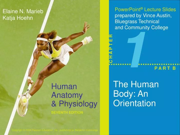
1. P A R T B. The Human Body: An Orientation. Anatomical Position. Body erect, feet slightly apart, palms facing forward, thumbs point away from body. Figure 1.7a. Directional Terms. __________________________– toward and away from the head, respectively

E N D
1 P A R T B The Human Body: An Orientation
Anatomical Position • Body erect, feet slightly apart, palms facing forward, thumbs point away from body Figure 1.7a
Directional Terms • __________________________– toward and away from the head, respectively • __________________________– toward the front and back of the body • __________________________– toward the midline, away from the midline, and between a more medial and lateral structure
Directional Terms • __________________________– closer to and farther from the origin of the body part • __________________________– toward and away from the body surface
Directional Terms Table 1.1a
Directional Terms Table 1.1b
Nasal (nose) Frontal (forehead) Oral (mouth) Orbital (eye) Buccal (cheek) Cervical (neck) Mental (chin) Acromial (point of shoulder) Sternal (breastbone) Axillary (armpit) Thoracic (chest) Abdominal (abdomen) Mammary (breast) Brachial (arm) Antecubital (front of elbow) Antebrachial (forearm) Umbilical (navel) Pelvic (pelvis) Carpal (wrist) Pollex (thumb) Palmar (palm) Coxal (hip) Digital (fingers) Inguinal (groin) Pubic (genital region) Femoral (thigh) Patellar (anterior knee) Fibular, or peroneal (side of leg) Crural (leg) Tarsal (ankle) Hallux (great toe) Pedal (foot) Digital (toes) (a) Anterior Regional Terms: Anterior View Figure 1.7a
Otic (ear) Cephalic (head) Occipital (back of head or base of skull) Acromial (point of shoulder) Vertebral (spinal column) Scapular (shoulder blade) Brachial (arm) Upper extremity Dorsum or dorsal (back) Olecranal (back of elbow) Lumbar (loin) Sacral (between hips) Manus (hand) Gluteal (buttock) Perineal (region between the anus and external genitalia) Lower extremity Femoral (thigh) Popliteal (back of knee) Sural (calf) Calcaneal (heel) Plantar (sole) (b) Posterior Regional Terms: Posterior View Figure 1.7b
Body Planes • _____________– divides the body into right and left parts • __________________________– sagittal plane that lies on the midline • __________________________– divides the body into anterior and posterior parts • __________________________ (cross section) – divides the body into superior and inferior parts • __________________________– cuts made diagonally
Body Planes Figure 1.8
Anatomical Variability • Humans vary slightly in both external and internal anatomy • Over _____________of all anatomical structures match textbook descriptions, but: • Nerves or blood vessels may be somewhat _____________ • Small muscles may be _____________ • Extreme anatomical variations are seldom seen
Body Cavities • Dorsal cavity protects the _____________, and is divided into two subdivisions • _____________– within the skull; encases the _____________ • _____________– runs within the vertebral column; encases the _____________ • Ventral cavity houses the internal organs (viscera), and is divided into two subdivisions • ________________ • ________________
Cranial cavity (contains brain) Thoracic cavity (contains heart and lungs) Dorsal body cavity Diaphragm Vertebral cavity (contains spinal cord) Abdominal cavity (contains digestive viscera) Key: Pelvic cavity (contains bladder, reproductive organs, and rectum) Dorsal body cavity Ventral body cavity (a) Lateral view Body Cavities Figure 1.9a
Cranial cavity Vertebral cavity Superior mediastinum Thoracic cavity (contains heart and lungs) Pleural cavity Pericardial cavity within the mediastinum Diaphragm Ventral body cavity (thoracic and abdomino- pelvic cavities) Abdominal cavity (contains digestive viscera) Abdomino- pelvic cavity Key: Pelvic cavity (contains bladder, reproductive organs, and rectum) Dorsal body cavity Ventral body cavity (b) Anterior view Body Cavities Figure 1.9b
Body Cavities • Thoracic cavity is subdivided into two pleural cavities, the mediastinum, and the pericardial cavity • Pleural cavities – each houses a _____________ • Mediastinum – contains the pericardial cavity; surrounds the remaining _____________ • Pericardial cavity – encloses the _____________
Body Cavities • The _______________________ is separated from the superior thoracic cavity by the dome-shaped _______________________ • It is composed of two subdivisions • Abdominal cavity – contains the _______________________ _______________________ • Pelvic cavity – lies within the pelvis and contains the _______________________ _______________________ ________________
Ventral Body Cavity Membranes • Parietal serosa lines _______________________ • Visceral serosa covers the _______________________ • Serous fluid separates the _______________________
Serous Membrane Relationship Figure 1.10a
Heart Serosae Figure 1.10b
Other Body Cavities • _______________________– mouth and cavities of the digestive organs • _______________________ –located within and posterior to the nose • _______________________– house the eyes • _______________________– contains bones (ossicles) that transmit sound vibrations • _______________________– joint cavities
Other Body Cavities Figure 1.13
Abdominopelvic Regions Figure 1.11a
Organs of the Abdominopelvic Regions Figure 1.11b
Abdominopelvic Quadrants • Right upper • Left upper • Right lower • Left lower Figure 1.12