Download
1 / 131
1.31k likes | 1.84k Views
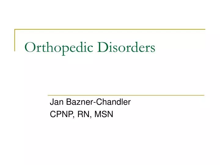
Orthopedic Disorders. Jan Bazner-Chandler CPNP, RN, MSN. Alterations in Musculoskeletal Status. Bowden & Greenberg. Musculoskeletal Differences in Children. Epiphyseal growth plate present Bones are growing / heal faster Bones are more pliable Periosteum thicker and more active

E N D
Orthopedic Disorders Jan Bazner-Chandler CPNP, RN, MSN
Alterations in Musculoskeletal Status Bowden & Greenberg
Musculoskeletal Differences in Children • Epiphyseal growth plate present • Bones are growing / heal faster • Bones are more pliable • Periosteum thicker and more active • Abundant blood supply to the bone • The younger the child the faster the healing.
Focused Physical Assessment • Inspect child undressed • Observe child walking • Spinal alignment • ROM • Muscle strength • Reflexes
Assessment Concerns: • Pain or tenderness • Muscle spasm • Masses • Soft tissue swelling
CoREminder • If an injury has occurred, examine that area last and be gentle when palpating the injury site
Nursing Alert • A child younger than 1 year who presents with a fracture should be evaluated for possible physical abuse or an underlying musculoskeletal disorder that would cause spontaneous bone injury.
Neurovascular Assessment • Pain • Where is it? • Is it reduced by narcotics? • Does the pain become worse when fingers or toes are flexed?
Neurovascular Assessment • Sensation • Can the child feel touch on the affected extremity • Motion • Can the child move fingers or toes below area of injury / nerve injury • Temperature • Is the extremity warm or cool to touch
Neurovascular Assessment • Capillary refill • Sluggish capillary refill may signals poor circulation • Color • Note color of extremity and compare with unaffected limb • Pulses • Assess distal to injury or cast
Neurovascular Impairment • Restriction of circulation and nerve function from injury or immobilizing device.
Clinical Manifestations • Increased pain • Edema • Decreased movement or sensation • Diminished or absent pulses distal to injury • Patient often described as restless – pain medication do not work – pain described as deep
Interventions • Assess area distal to injury, cast, splint, traction for adequate circulation • Release pressure by splitting the cast or loosening restrictive bandage. • Notify physician
Compartment Syndrome • Pain is the hallmark sign, pain out of proportion to the normal clinical course. • Must be diagnosed immediately or irreversible neurovascular, muscular, vascular damage occurs that can lead to renal failure and death.
Clinical Manifestations • A combination of signs and symptoms characterize compartment syndrome. The classic sign of acute compartment syndrome is pain, especially when the muscle is stretched. • There may also be a tingling or burning sensation (paresthesias) in the muscle. • A child may report that the foot / hand is “a sleep” • If the area becomes numb or paralysis sets in, cell death has begun and efforts to lower the pressure in the compartment may not be successful in restoring function.
Physical Assessment • Frequent pain assessment • If pain med does not work something is wrong • The muscle may feel tight or full. • Measure the affected muscle group and compare with the unaffected side • Pulses below area of injury
Treatment • Prevention • Don’t elevate the affected limb above or below the level of the heart. • Dressings should be removed if CS is suspected. • Casts should be bi-valved in high risk situations.
Assessment • Don’t forget the five P’s • Pain • Paresthesia • Passive stretch • Pressure • Pulse-less-ness
Surgical Management Siumed.edu Fasciotomy to relieve pressure. The fascia is divided along the length of the compartment to release pressure within.
Nerve Assessment • Important to due on admission from ER or to the unit • Repeat after cast, traction, or surgery done on the extremity
Treatment Modalities • Goals of fracture care: • To regain alignment and length of the bony fragments • To retain alignment and length • To restore function of the injured part
Traction • Realign bone fragments • Provide rest • Prevent or improve deformity • Pre or post operative positioning • Reduce muscle spasm • immobilization
Fractures Treatment determined by type of fracture
Fractures RW Chandler MD
Salter-Harris Classification If injury involves growth plate in an immature bone, growth disturbance may follow. • Classification system describes the injury and the potential for growth disturbance.
Bucks Traction Ball & Bindler
Principles of Traction • Counter traction with weights • Make sure all ropes and pulleys are aligned and weights are hanging freely • Do not remove weights unless instructed to do so • Traction must be applied at all times
Bryants Traction • Used for child under3 yrs • Hip dysplasia / fractured femur • Buttocks do not rest on mattress • Assess neurovascular and restriction by ace bandages – compartment syndrome
Skeletal Traction • Pull directly applied to bone by pin • Pin care • Increased risk of infection Ball & Bindler
External Fixation RWChandler MD
External Fixator Ball & Bindler
Pin Care • Provide pin care as ordered. Cleanse area around pin with normal saline or half-strength hydrogen peroxide. • Have parent / caretaker demonstrate pin care before discharge
External Fixator RW Chandler MD
External Fixator RW Chandler MD
Plates and Pins Plates, screws, and wires are used to align bone fragments. R.Chandler MD
Post-operative Care • Assess color, sensation, cap refill, movement, pain, and pulses • Circle any drainage noted on cast or dressing. • Pain control • Edema = ice to area • Pulmonary function = C&DB
Pulmonary Embolism • A complication of a fractured leg is a pulmonary embolism. Fat escapes the marrow when the bone is fractured and can travel through the blood stream and become lodged in small vessels like the arterioles and capillaries of the lung. • Primary symptom is shortness of breath and chest pain.
Interventions • Place patient in high fowlers • Administer oxygen • Call MD • Chest x-ray • Outcomes are better for a health person; poorer for person with pre-existing lung problems.
Orthopedic Disorders • Congenital • Acquired / trauma • Infectious
Tales Equinovarus Tales equinovarus or Club foot Obvious deformity noted at birth. Surgical correction Bowden & Greenberg
Tales Equinovarus • Club Foot • 1 to 2 per 1000 • Males more affected • Involves both the bony structures and soft tissue. • The entire foot is pointing downward.