Download
1 / 122
1.22k likes | 1.49k Views
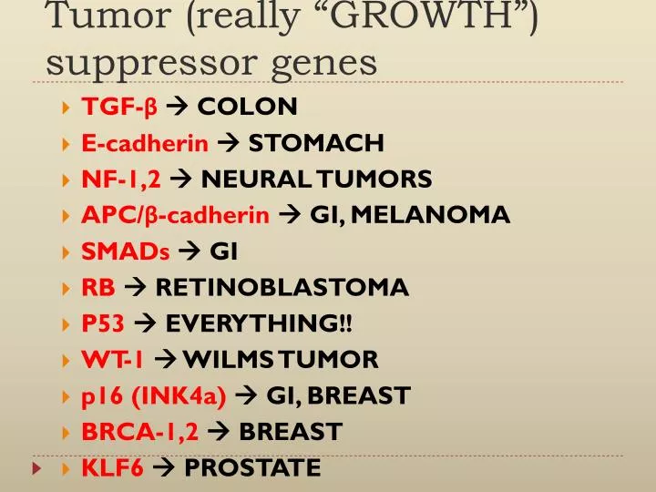
Tumor (really “GROWTH”) suppressor genes. TGF- β COLON E- cadherin STOMACH NF-1,2 NEURAL TUMORS APC/ β - cadherin GI, MELANOMA SMADs GI RB RETINOBLASTOMA P53 EVERYTHING!! WT-1 WILMS TUMOR p16 (INK4a) GI, BREAST BRCA-1,2 BREAST KLF6 PROSTATE.

E N D
Tumor (really “GROWTH”) suppressor genes • TGF-β COLON • E-cadherin STOMACH • NF-1,2 NEURAL TUMORS • APC/β-cadherin GI, MELANOMA • SMADs GI • RB RETINOBLASTOMA • P53 EVERYTHING!! • WT-1 WILMS TUMOR • p16 (INK4a) GI, BREAST • BRCA-1,2 BREAST • KLF6 PROSTATE
TRANSFORMATION & PROGRESSION • Self-sufficiency in growth signals • Insensitivity to growth-inhibiting signals • Evasion of apoptosis • Limitless replicative potential: Telomerase • Angiogenesis • Invasive ability • Metastatic ability • Defects in DNA repair: “Spell checker”
Evasion of APOPTOSIS • BCL-2 Overexpression • Seen in B-cell lymphomas of follicular type t(14;18)(q32;q21),dominant with IgH genes and overexpression of BCL-2 protein • P53 (Pro-apoptic gene) - Directly activates Bax (Bcl-2 protein) • Reduced levels of CD95/Fas • FLIP protein – Inhibits Caspase 8 • Loss of APAF1(Apoptosis activating factor-1) • Increased IAP (inhibitors of apoptosis)
LIMITLESS REPLICATIVE POTENTIAL • TELOMERES determine the limited number of duplications a cell will have! • TELOMERASE, present in >90% of human cancers, changes telomeres so they will have UNLIMITED replicative potential
TUMOR ANGIOGENESIS • Q: How close to a blood vessel must a cell be? • A: 1-2 mm • Activation of VEGF and FGF-b • Tumor size is regulated (allowed) by angiogenesis/anti-angiogenesis balance
Proteases of tumors can induce production of angiogenic factors and inhibitors • Proteases act on ECM components (E.g. Collagen) releasing bFGF and VEGF • VEGF triggers the NOTCH Pathway
INVADE & METASTASIS • 1. Invasion of Extracellular Matrix • 2. Vascular Dissemination • Invading the Extracellular matrix requires breaching the basement membrane Intersitial Connective Tissue Vascular Basement Membrane
Invasion of ECM • 1. Detachment ("loosening up") of the tumor cells from each other • Down-regulation of E-cadherin and Beta-catenin reducing cell’s adherance to each other
Invasion of ECM • 2. Degradation of ECM, e.g., collagenase, etc. • Break down the basement membrane • Proteases – MMP (matrix metalloproteinases) • Releasing growth factors (e.g. VEGF) and angiogenic, growth promoting effects with cleavage of Type IV collagen for example • 3. Attachment to matrix components • Breakdown by MMP allows tumor cell receptors to bind ECM proteins and stimulate migration • E.g. MMP2 cleaves laminin and MMP9 cleaves collagen IV Change in tumor cell receptor attachment to ECM proteins Stimulates Migration
Invasion of ECM • Migration of tumor cells • Cytokines ( autocrine factors) • Cleavage of the ECM components (Laminin, collagen) and growth factors (IGF I, II) that have chemotactic activity • Stromal cells around tumor also producehepatocyte growth factor-scatter factor promoting motility
TRANSFORMATIONGROWTHBM INVASIONANGIOGENESISINTRAVASATIONEMBOLIZATIONADHESIONEXTRAVASATIONMETASTATIC GROWTHetc.
Vascular Dissemination • Following intravasation, clumps of platelet-tumor aggregates are formed • Extravastion of this tumor emboli at distant sites Attachment to the endothelium Exits through the basement membrane • Followed by migration to lymphoid tissue using the CD44 adhesion molecule (expressed on T cells) • CD44 binds to hyaluronate allowing migration and towards metastic spread • *Most metastases occur in the first capillary bed available to tumor
Other possible mechanisms explaining metastasis: • 1. Tumor cell adhesion molecule ligandsare expressed on the endothelial cell of target tissue • Target organ variation in endothelial cell expression of adhesion molecule ligands • 2. Chemokinereceptors are expressed in cancer cells that allowing binding of chemokines expressed at specific tissues • E.g. CXCR4 and CCR7 receptors expressed in breast cancer cells • 3. Chemoattractants – E.g. IGF 1, II, can be expressed at target tissues
Breast cancer cells, how do they adapt and make their environment habitable? • - Osteolytic behavior in bone by osteoclasts • Tumor cells PTHRP (parathyroid hormone-related protein) Osteoblasts RANK ligand Osteoclasts • Osteoclasts break down the matrix releasing growth factors
Development of Metastasis • One hypothesis suggest why only some tumors metastasize? • “Metastasis Signature” Multiple abnormalities taking place in the early stage of development • Intrinsic properties, microenvironment (stroma), angiogenesis, resistance to immune defenders, and local invasiveness • Other hypothesis Rare clonal variation that develop metastasis
DEFECTS IN DNA REPAIR • 1. Mismatch Repair • 2. Nucleotide Excision Repair • 3. Recombination Repair
DNA REPAIR GENE DEFECTS • DNA repair is like a spell checker • HNPCC (Hereditary Non-Polyposis Colon Cancer [Lynch]): TGF-β, β-catenin, BAX • XerodermaPigmentosum: UV fixing gene • Ataxia Telangiectasia: ATM gene • Bloom Syndrome: defective helicase • Fanconi anemia
Hereditary Non-PolyposisColon Cancer • Mismatch Repair • Proofreaders disabled • Microsatellite fragments 1 to 6 nucleotide repeats • Creates instability • Dna mismatch repair gene affect cell growth indirectly, through mutations in other genes TGF-beta, beta-catenin, Bax
XerodermaPigmentosum • UV radiation exposure • Pyrimidinedimers • DNA damage with defect in nucleotide excision repair
Autosomal Recessive Disorders • Defect in DNA repair by Homologous Recombination • Ataxia – Telangiactasia– neurological degenerative disorder, exposure to Ionizing radiation • Defective ATM gene • Bloom Syndrome - Ionizing radiation resulting in developmental defects • Defect in gene of chromosme 15 encodes helicase dna repair through homologus recombination • Fanconi anemia – Chemotherapeutic agents causing damage • Bone-marrow aplasia BRCA1 and BRCA 2 Proteins take part in homologous recombination repair pathway • Mutation of BRCA2 gene chromosomal breaks & aneuploidy
STROMAL MICROENVIRONMENT • “Cross-Talk” between the tumor cells and ECM • E.g. Laminin degradation by MMP2 Stimulates tumor cell motility • E.g. Type IV collagen degradation by MMP9 Release of VEGF • Storage of inactive growth factors in ECM, proteases release the active form (E.g. PDGF, TGF-beta, bFGF)
STROMAL MICROENVIRONMENT • Inflammatory Cells and fibroblasts • Cytokines released during inflammation Induce survival and progression of tumor cells • Chemokines Chemotactic properties • Fibroblasts Important role in tumor cells • Desmoplastic response, highly fibrotic , promoting growth
WARBURG EFFECT • Otto Warburg, Nobel Prize discovery on the metabolism of turmors • Cancer cells rely on Aerobic Glycolysis, Warburg Effect, instead of the mitochondrial oxidative phosphorylation • Several Hypothesis – • 1. During angiogenesis, vessels are poorly formed, activation of H1F1alpha due to hypoxia increases expression of enzymes of glycolysis, and vasculature • 2. Exposure to state of continous hypoxia and normoxia • 3. Mutations in oncogene and tumor supressors (p53, RAS, PTEN) stimulate metabolic changes
Metabolic Alterations • In addition, tumor cells need to build and maintain its membrane, proteins, and organelles --- Anabolic Pathway • Lipid, nucleotide, proteins (amino acids), glucose (carbon) • Glutamine Both glycolytic and anabolic pathway
PI3K/AKT/Mtor pathway • Important in regulating normal cell functions including amino acid, glucose uptake • In tumors, mutation of tumor supressors and oncogenes --- Abnormality in pathway can occur favoring survival and proliferation of tumor cells • E.g. PTEN has a negative regulation of this pathway,
Other tumor supressors that supress this pathway:- • Peutz-Jegher syndrome mutated gene LKB1 ( Liver kinase B1) • – Limits or regulates cell growth by AMP-dependent protein kinasemechanism – Negative regulator of mTOR pathway • Supression of LKBI (also called STK11) Increase in anabolic metabolism of cancer cells • TuberousSclerosis mutated genes (TSC1 and TSC2) ----- Negative regulation of mTOR
CHROMOSOME CHANGES in CANCER • TRANSLOCATIONS and INVERSIONS • Occur in MOST Lymphomas/Leukemias • Occur in MANY (and growing numbers) of NON-hematologic malignancies also
Translocations ------------- Activate Proto-Oncogenes • 2 common mechanism of translocation: • A) Lymphoid Tumors --- Swapping or movement of segment of chromosome to another chromosome • B) Hematopoietic tumors --- Two chromosomes recombine to form hybrid fusion genes for production of chimeric proteins
Burkitt Lymphoma:- t(8:14) (q24;q32) • Translocation at chromosome 8 (c-MYC) and 14 ( IGH gene) • MYC containing segment of chromosome 8 movement to to chromosome 14q32 at IGH gene • Overexpression of MYC
Follicular Lymphomas:- t(14:18) (q32:q21) • Translocation at chromosome 14 (IGH gene) and 18 (BCL2) • Activation of BCL2
Chronic Myeloid Leukemia (CML):- t(9;22) (q34;q11) • Translocation at chromosome 9 (ABL gene) and 22 (BCR gene)
What is the Philadelphia chromosome? • Chromosome 22 that exchanges portion (reciprocal translocation) of the chromosome with Chromosome 9 • Fusion of two genesBCR-ABL producing protein with tyrosine kinase activity • Found in both CML & ALL (Acute Lymphoblastic Leukemia)
Ewing Sarcoma • Also known as PNET (Primitive neuroectodermal tumor) • T(11:22) (q24;q12) FLI 1 gene and EWSR1 gene • Chimeric protein EWS-FLI 1 • Fusion of most genes encode for transcription factors
GENE AMPLIFICATION • Amplification of particular chromosomal regions that contain the oncogenic sequence • Hallmark Features:- • Double minutes ---- Small, centric structures or chromatin bodies of extrachromosomaldna • Homogenous staining regions • Once a gene is amplified, these amplified regions can be inserted into new choromsome • Lack a normal banding pattern and appear homogenous (G-banded karyotype)
Gene Amplification • N-MYC ---- Neuroblastoma • ERBB2 --- Breast Cancers • C-myc, L-myc, N-myc --- Small cell lung cancer • **Greater the amplification, poorer the prognosis
Epigenetic Changes • Heritable changes in gene expression without changing the DNA sequences • Post-translational modfications of histones+ DNA methylation ===== Gene Expression • Cancer cells --- Hypermethylation within promoter regions and global DNA Hypomethylation • Tumor supressor genes --- hypermethlation --- Silenced • E.g. BRCA1, VHL, MLH1
Epigenetic Changes • Genomic Imprinting: • Maternal or paternal allele of a gene or chromosome is modified by methylation and inactivated
miRNAs • Non-coding, single stranded RNA • RNA-induced silencing complex • Post-transcriptional gene silencing • Downregulate gene expression by translational repression, and deadnylation • In cancer cells, increase oncogene expression or reduce tumor supression gene expression
miRNAs • 1. Reduced miRNA (Inhibitor of oncogene translation) --------------- Overproductin of oncogene • 2. miRNA reduces tumor supressor gene