Download
1 / 37
370 likes | 549 Views
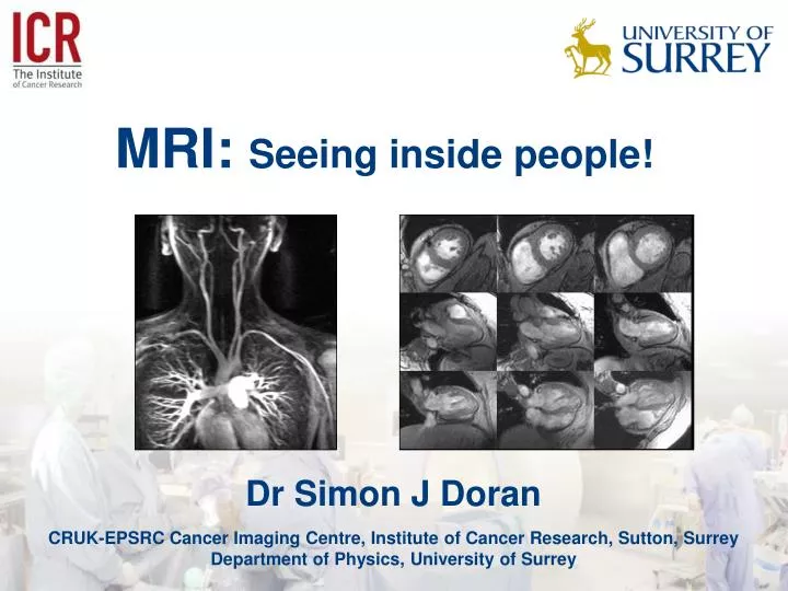
MRI: Seeing inside people!. Dr Simon J Doran CRUK-EPSRC Cancer Imaging Centre, Institute of Cancer Research, Sutton, Surrey Department of Physics, University of Surrey. Summary of today’s talk. Brief history of medical imaging A few basic principles The human brain as seen by MRI

E N D
MRI: Seeing inside people! Dr Simon J Doran CRUK-EPSRC Cancer Imaging Centre, Institute of Cancer Research, Sutton, Surrey Department of Physics, University of Surrey
Summary of today’s talk • Brief history of medical imaging • A few basic principles • The human brain as seen by MRI • What else can MRI do?
History of medical imaging • There is a lot of evidence that the ancient Greeks performed major surgery as early as 150 BC. • But how did they know what was inside the body?
History of medical imaging • Up until the very end of the 1800’s, the only way to find out about what was inside the human body was to find somebody who was already dead and cut them up. “The Anatomy Lecture of Dr. Nicolaes Tulp” [1632] by Rembrandt • This is not very useful if you want to find out what is wrong with somebody who is alive and cure them!
History of medical imaging Mrs Roentgen’s hand Painting of woman having an x-ray (1896) Modern x-ray Data: Mayo Clinic • Things changed for ever in 1895 with the discovery of X-rays by Wilhelm Roentgen.
History of medical imaging • The problem with x-ray imaging is that you can only see flat 2-D images. • By shining x-rays through the body at a variety of different angles all round the body, we can reconstruct images that are three-dimensional. Sir Godfrey Hounsfield and his x-ray CT scanner • In 1972, Sir Godfrey Hounsfield made the breakthrough that earned him the Nobel Prize.
History of medical imaging Data :Toshiba AmericaVisuals : Vital Images X-ray CT visualisation: Vital Images • X-ray CT is particularly good for seeing bones and blood vessels.
History of medical imaging Data source : SMIS Ltd Sir Peter Mansfield and Paul Lauterbur, Photo: AFP • Just two years later (1974), these two men invented the MRI scanner • They got the Nobel Prize, too, but they had to wait till 2003. • MRI is really good at taking pictures of the brain.
Data: Mayo Clinic Data : Toshiba Visuals : Vital Images Why use different methods of imaging? • Plane-film X-ray maps the total attenuation of X-rays along a path through the body, giving a projection image. Good for bone structure in accidents. • X-ray CT measures the X-ray attenuation coefficient of the body at each point. True 3-D images. • Ultrasound maps the reflectionand attenuation of sound. Data: Clearview Ultrasound
Data source: SMIS Ltd Data: CSUA, Berkeley Data: FORENAP, Rouffach Why use different methods of imaging? • MRI maps the distribution and “environment” of water molecules in the body. • PET maps the distribution of radioactively labelled compounds. • MEG maps directly the magnetic fields generated by currents flowing in the brain.
What is the tunnel into which the patient slides? Image sources : GE Medical Systems, VA Imaging Centre, University of Florida
B Head Feet x What happens in a scan? • Resonant frequency is related to the magnetic field. • f B • If we vary the magnetic field across the sample, then the frequency of emitted radio waves varies. • By looking at the frequency spectrum of the signal, we can find out how many spins are where.
Why do MRI scanners make a noise? • A gradient coil is just like a big loudspeaker (i.e., a large coil of wire sitting in a magnetic field). • As we change the current passing through the coil the whole gradient assembly (many tons) tries to move. • It is held in place very securely, yet still vibrates a little, particularly at certain frequencies. • An expert can tell what imaging scan is being done by listening to the sound the gradients make. Scout T1-W gradient echo EPI
The Human Brain as seen by MRI Data: The Whole-brain Atlas, K. A. Johnson and J. A. Becker, Harvard Data: Christopher Nimsky, Neurosurgery, Erlangen, Germany
Where are we heading with anatomical MRI? 7 T Data source: SMIS Data source: Gachon University, Seoul
Data source: Roger Ordidge, UCL Where are we heading with anatomical MRI? 4.7 T Stained section 1.5 T
MRI in research: Functional imaging • We acquire one image of the brain in a “resting” state. • We follow this by a corresponding image where the brain is active. • Any differences between these two images correspond to places where the brain is working. Data source: http://www.youtube.com/watch?v=alS3GeRxYGY • We can see you think!!
MRI in research: Functional imaging • Typical base data (greyscale) at 4.7 T, leading to the activation time course (right) and subsequent overlaying of activated areas on images (yellow-red scale) Data source: Roger Ordidge, UCL
Nerve fibre MRI in research: neural fibre tracking • MR images can be sensitised to the rate of diffusion of water molecules. • Water diffuses faster along nerve fibres than perpendicular to them. • This allows us to map the local direction of a fibre and create a map of the fibres. Data source: Geoff Parker, University of Manchester
MRI in research: neural fibre tracking • MR images can be sensitised to the rate of diffusion of water molecules. • Water diffuses faster along nerve fibres than perpendicular to them. • This allows us to map the local direction of a fibre and create a map of the fibres. • Finally, we can overlay them on a high-resolution 3-D image of the head. Data source: Wilde et al. 2008
MRI in research: neural fibre tracking Data source: Wakana et al., Radiology 2004; 230:77–87
MRI in research: neural fibre tracking Data source: András Jakab. (University of Debrecen)
MRI-guided neurosurgery • State-of-the-art neurosurgery unit at Erlangen, Germany. • MRI is completely integrated into the operating theatre and used in conjunction with digital images captured from an operating microscope. Data source : Christopher Nimsky, Neurosurgery, Erlangen, Germany
MRI-guided brain surgery • The surgeon can see both the patient and MR image on the same display. Data source : Christopher Nimsky, Neurosurgery, Erlangen, Germany
MRI-guided thermotherapy • Basic premise: Heating tumour tissue in vivo can destroy tumours by “cooking them” • The basic requirements of the system are:. • a heating device • a method of monitoring the heating in 3-D • a method of controlling the heating based on feedback from the monitoring Data source : Chrit Moonen, University of Bordeaux, France
f z x MRI-guided thermotherapy • The MR signal can be made sensitive to temperature, allowing a temperature map to be made over a 3-D region in a matter of seconds. • The heating source is a focused ultrasound transducer, built into the scanner’s patient table. Data source : Chrit Moonen, University of Bordeaux, France
MRI-guided thermotherapy Colours indicate temperature distribution Muscle Water Transducer MR image with temperature map overlaid Planned temperature change, with measured values Data source : Chrit Moonen, University of Bordeaux, France
Ø = 2 mm Data source : Stefan Petersson, Malmö, Sweden MRI-guided surgery: catheter placement Schematic of homebuilt catheter Realisation of catheter
Data source : Stefan Petersson, Malmö, Sweden MRI-guided surgery: catheter placement 13C MR guided renal intervention TrueFISP pulse sequence (linear) TR/TE/FA 5.1 ms / 2.6 ms / 70o Pixelsize 2.0 x 2.0 x 200.0 mm3 Matrix 64x 128 Scan time 300 ms / projection Off-line reconstruction
MRI in research: Stem cell imaging cell Iron-oxide nanoparticles Unlabelled stem cells factor 50 higher iron content Magnetically labelled cells Data source: Peter Jakob, University of Wurzburg, Germany
MRI can image much more than just the brain ... Data source : Siemens Medical
MRI can image much more than just the brain ... Data: www.journey-with-crohns-disease.com Data: David Lomas, Addenbrokes Hospital A virtual tour of the human colon ...
Sports injuries ... Data: www.imaios.com/en/e-Anatomy/Limbs/Knee-MR Data: Paul Debevec
… and the diagnosis, Mr Beckham ... Data source: American Radiology Services
Conclusion • There are many different ways of imaging the human body. • The different methods tell us different things. • It is study of basic Physics (electromagnetism, nuclear physics, mechanics) which has discovered the principles. • It is money — the human brain is a very valuable thing — which has led to the incredible developments that we see today.
The End Thank you for listening! Any questions?