Download
1 / 27

E N D
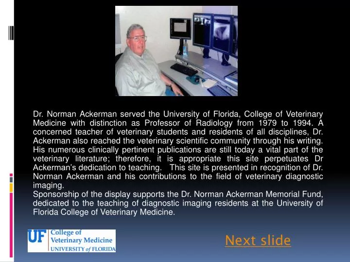
Dr. Norman Ackerman served the University of Florida, College of Veterinary Medicine with distinction as Professor of Radiology from 1979 to 1994. A concerned teacher of veterinary students and residents of all disciplines, Dr. Ackerman also reached the veterinary scientific community through his writing. His numerous clinically pertinent publications are still today a vital part of the veterinary literature; therefore, it is appropriate this site perpetuates Dr Ackerman’s dedication to teaching. This site is presented in recognition of Dr. Norman Ackerman and his contributions to the field of veterinary diagnostic imaging. Sponsorship of the display supports the Dr. Norman Ackerman Memorial Fund, dedicated to the teaching of diagnostic imaging residents at the University of Florida College of Veterinary Medicine. Next slide
Norman Ackerman Memorial Radiography Case Challenge • SAM • 9 year old MN Mixed Breed Dog Next Slide
Signalment • Sam presents to your clinic with a acute history of cough and exercise intolerance • On physical examination, you hear crackling lung sounds cranially, on the right side • You order thoracic radiographs Next Slide
Previous Slide Next Slide
Next Slide Previous Slide
Previous Slide Next Slide
Based on your assessment of the radiographs, the thoracic body wall is: • Normal • Abnormal Previous Slide
Correct! There are no abnormalities associated with the thoracic wall. Next Slide
Sorry! The thoracic body wall, including the extrathoracic structures, are within normal limits Click here to proceed to the next question
Based on your assessment of the radiographs, the pleural space is: • Normal • Abnormal
Correct! There are no abnormalities associated with the pleural space. Next Slide
Sorry! The pleural space is normal Click here to proceed to the next question
Based on your evaluation, the cardiac silhouette is: • Normal • Abnormal
Sorry, Try Again The cardiac silhouette is within normal limits. Click here to continue
Correct! There are no abnormalities associated with the cardiac silhouette Next slide
Based on your assessment of the radiographs, the lungs, including the vessels, are: • Normal • Abnormal
Sorry! • There is an abnormality associated with the lungs. Continue
Correct! There is an area of increased soft tissue opacity mainly on the ventral aspect of the right cranial lung lobe. Based on your assessment, which pulmonary pattern is predominant within that lobe? • Bronchial • Alveolar • Vascular • Unstrutured Interstitial
Sorry! Indefinition of the pulmonary vessels, air bronchograms, lobar sign, and (in this case, discrete) border effacement of the lobar opacification with the cardiac silhouette are not characteristics of this pulmonary pattern Previous Slide
Correct! This is an example of an alveolar pulmonary pattern. Some of the features of this pattern include: Indefinition of the pulmonary vessels, air bronchograms, lobar sign, and (in this case, discrete) border effacement of the lobar opacification with the cardiac silhouette. Remember: It does not have to have all these features to be considered an alveolar pattern! Continue
LS AB IV BE AB=air bronchogram IV=indefinition of vessels BE=border effacement on the cardiac silhouette LS=lobar sign Continue Previous Slide
Conclusion Your findings now include: increased soft tissue pulmonary opacity within the right cranial lung lobe, with presence of indefinitionof the pulmonary vessels, air bronchograms, lobar sign, and (in this case, discrete) border effacement of the lobar opacification with the cardiac silhouette. This represents an alveolar pulmonary pattern, which, in this case, is mainly ventral. click next.
Conclusion • What is top differential diagnosis? • Cardiogenic pulmonary edema • Aspiration pneumonia • Recumbence atelectasia
Sorry! • Usually cardiogenic edema has a caudodorsal distribution within the lung parenquima of dogs • Although we cannot totally ruled out cardiac disease just using radiographs, the cardiac silhouette is within normal limits in this case. One more try!
Sorry! • In addition to the soft tissue opacification within the lungs, in cases of atelectasis, usually is observed a decreased volume of the affected lung lobe, and sometimes ipsilateralmediastinal shift (MS). In Sam’s case, the volume of the right cranial lung lobe is normal (not decreased). MS One more try!
Correct! • Aspiration pneumonias are usually ventral, due to gravitational forces. • This also goes along with the acute clinical signs. Next
Some causes of Aspiration Pneumonia (Dennis, Kirberger, Barr, Wrigley: Handbook of Small Animal Radiology and Ultrasound, 2nd ed., 2010): • Regurgitation and vomiting, especially if esophageal dilation is present; • Iatrogenic aspiration: force feeding, medication, anesthesia and oral administration of contrast medium; • Swallowing disorders; • Weakness and debilitation; • Cleft palate; • Tracheo-esophageal or broncho-esophageal fistula. Return to the webpage Return to the beginning