Download
1 / 31
590 likes | 1.58k Views
Bioseparation Engineering. Protein Purification Processes. Protein Purification Processes. Final product requirement – define (99% ~ 99.999%). Process design approaches. Follow examples Modify the process (new concept can be introduced).

E N D
Bioseparation Engineering Protein Purification Processes
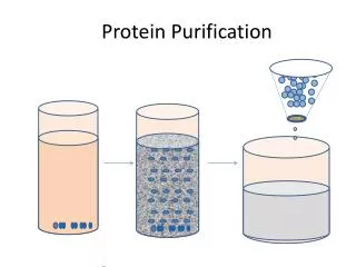
Protein Purification Processes Final product requirement – define (99% ~ 99.999%) Process design approaches • Follow examples • Modify the process • (new concept can be introduced) Based on different physical, chemical, biochemical properties Separate the most plentiful impurities, first Differences in physicochemical properties High resolution Most difficult step, last Rule
Protein Purification Processes Cell separation Cell disruption, debris separation (for intracellular protein) Concentration High resolution purification Polishing of final product Efficiency: 0.9 for each step is assumed after 6 ~ 7 steps → total yield ≈ 0.2 • increase yield/efficiency is very essential • refolding is important
Protein Extracellular protein (secreted): easy to separate Inclusion body: cell disruption → centrifuge solubilization → refolding - - chromatography Inclusion body is formed by overexpression in E. coli. Advantages of Inclusion Body • not breakable with protease • not toxic to cells • high expression In general, economical
What to remove? • cell debris, endotoxin, other proteins, nucleic acid • deaminated form, oxidized form, dimer form of the product protein • need more than 3 steps of chromatography
Protein Purification Processes 1. Cell Harvest by Centrifugation • Continuous disk stack type or batch centrifuge • tangential flow microfiltration (M/F) 2. Cell Disruption • French press, lysozyme, ultrasonification or bead mill E. coli: mainly high pressure homogenizer 2 – 3 times for high efficiency Yeast: strong cell wall → bead mill
3. Solubilization of Inclusion Body Inclusion body: can be obtained at 60 – 70% purity Denaturing Agent (chaotropic agent) Urea, guanidine HCL, NaOH (high pH) In case of urea, it should have no cyanate. protein + cyanate → carbamylation occurs Guanidine: good but expensive So, 7 – 8 M urea is widely used. If disulfide bond exists in protein, add mercaptoethanol, DTT (dithiothreitol) or cysteine → reduce disulfide bond to free thiol
Add 1-2 mM EDTA to reduce metalloprotease activity. After solubilization, viscous solution containing the following is obtained. 70% of target protein, cell debris, DNA, endotoxin, 7 – 8 M urea, 50 – 100 mMcystein, 1 – 2 mM EDTA • Host which has no protease is used. • Short processing time is required.
4. Capture Impurities For large volume, short time → chromatography If histidine tag exists → IMAC (immobilized metal affinity chromatography) If no histidine tag → urea → ion-exchange chromatography After this process, solution having the following is obtained. target protein (1 – 5 mg/ml), small impurities, 7 – 8 M urea, 50 – 100 mMcystein, 1 – 2 mM EDTA, pH 7.9 – 9.0 buffer
5. Refolding • Remove denaturant • method: dialysis, diafiltration • Refolding is being performed at low concentration (0.1 – 0.5 g/L) to prevent protein aggregation 6. Purification • IEC, IMAC, HIC • HIC: hydroxyapatite chromatography • diafiltration or gel permeation chromatography for desalting (to exchange buffer solution)
7. Polishing • Remove dimer, polymer, endotoxin • Use chromatography • After polishing → filtration using 0.2μ filter to remove bacteria • Protein from mammalian cell • - secreted form • inactivate virus, virus removal process is required • To prevent protein oxidation • operation under nitrogen gas • low temperature is preferred
Evaluation of Separation and Purification Processes in the Antibiotic Industry 2011-21046 Yang jinkyoung
Conventional separation technologies • Two main processing segments; • fermentation section • separation and purification section • All antibiotic fermentations use similar equipment, while different antibiotics are produced by using different cultures and growth media. • The separation and purification sections of an antibiotic plant can differ substantially depending on the specific antibiotic that is being produced and enduse purity requirements. • Filtration, centrifugation, extraction, and crystallization are generally employed.
Penicillin • With proper strain selection penicillin can be produced in concentrations up to 40 g/L, which is one or two orders of magnitude greater than many other antibiotics. • Cephalosporin • broad-spectrum antibiotics that have low toxicity. • They are produced by the same process used for the penicillins, utilizing a different growth medium and organisms. • The final fermentation broth concentrations are one to two orders of magnitude lower than penicillin, resulting in a more difficult separation and purification process.
Recovery of cephalosporin from the filtrate is difficult because of the low product concentration and the need to remove high molecular weight biological compounds. • During biosynthesis of CPC, the formation of the synthesizing enzyme is sequentially induced in the metabolic pathway. It is therefore necessary to separate/isolate the enzymes. (CPC)
General production scheme for cephalosporin • Many different separation and purification schemes are employed, including conventional solvent extraction, ion exchange resins, and salting out procedures. Adding water and a polar organic solvent Anion exchange Fermentation Adsorption W/ active C Filtration Adding salt solution • Frequently, the carbon adsorption steps are replaced with a precipitation step. Many different precipitations are possible. • 1. Crystallization of the potassium or sodium salt from purified aqueous solution of the cephalosporin by concentration and/or addition of large volumes of a miscible solvent. • 2. The zinc salt (also copper, nickel, lead, cadmium, cobalt, iron, and manganese) can be crystallized from purified aqueous solutions. • 3. Insoluble derivatives such as the n-2,4-dichlorobenzoyl cephalosporin and tetrabromocarboxybenzoyl cephalosporin are crystallized as the acid from solution. • 4. Sodium-2-ethyl hexanoate will precipitate the sodium salt of N-derivatizedcephalosporins from solvents.
Isomerization This is biologically inactive.
Difficult to isolate and purify due to a highly polar side-chain.
Example 10.7 Validation of the preparation of clinical monoclonal antibodies 2011-21071 임준혁
Briefly describe the validation procedure to purify monoclonal antibodies (MAbs) from mouse ascites fluid (Mariani & Tarditi, 1992) • Ammonium sulfate precipitation • Two-step HPLC method • Protein A chromatography • Hydroxyapatite chromatography • 4.5 mg of IgG in 0.5 mL murine ascites with 4 different viruses (representative contaminating agent) • Eluent : buffer
Elimination of Viruses during MAb purification Ammonium sulfate precipitation ÷ 102.91 Protein A chromatography Hydroxyapatite chromatography
Elimination of Murine DNA during MAb purification FDA recommendation : below 10 pg per dose (of 1mg) Initial concentration of the DNA : 5~10 ng/mg Final concentration of the DNA : 5~10 fg/mg
Elimination of protein A from the final product FCR recommendation : below 1 ppm per dose After protein A chromatography : 40-ppm protein A at most → hydroxyapatite chromatography should reduce protein A by a factor of 50 The protein A level in the final product : 0.24 ppm
Two-step HPLC method • adsorbing the protein to Protein A (Hydroxylapatite) immobilized on a solid phase comprising silica or glass • Protein A • Protein A is a 40-60 kDa MSCRAMM surface protein originally found in the cell wall of the bacterium Staphylococcus aureus. • Protein A binds with high affinity to human IgG1 and IgG2 as well as mouse IgG2a and IgG2b • Hydroxylapatite • a naturally occurring mineral form of calcium apatite (Ca5(PO4)3(OH)) • "mixed-mode" ion exchange • nonspecific interactions between positively charged calcium ions and negatively charged phosphate ions
2011-2 Prof. Young Je Yoo Bioseparation Engineering Example 5.9 Provide an example for heparin HPLC separation of proteins 2011. 11. 7 JongHoon Kim
Paper. Affinity high-performaceliqiud chromatography on heparin-Glc-Spheron • Heparin • Sulfuric ester of a complex carbohydrate containing glucosamine glucuronic acid • Various biological function • -Selective binding with antithrombinIII • → increase inhibitory activity • -Helps in inactivating other vitamin K-dependent coagulation factor • -Polyanionic nature : interaction with cationic biological compounds →purification by affinity chromatography • Affinity chromatography could serve as an analytical tool to detect and measure the fuctional binding characteristics • immobilize heparin on a rigid high-performance affinity support
Two methods of heparin immobilization on Glc-Spheron HGS I ( Heparin-Glc-Sepharon I) ① gel was suspended in 1M NaOH , activated with epichlorhydrin (60℃, 2hr) ② Cooling , washing with distilled water ③ coupling heparin at pH 9.0 (borate buffer, room temp, 48hr) - the amount of immobilized heparin : 4mg/ml of the gel 2. HGS II (Heparin-Glc-Shpharon II) ① gel was activated with 1M sodium peroxidate ( room temp, 2hr) ② immobilizing heparin at pH 6.0 (12hr) ③The bods were stabilized by reduction with sodium borohydride(pH 9.0) to increase the chemical stability of the bond - the amount of immobilized heparin : 1mg/ml of the gel
Result and Discussion I. Antithrombin III - Highest affinity from plasma proteins → capacity of HGS I : 2mg/ml HGS II : 1mg/ml - Change in the flow-rate from 1ml/min to 0.5ml/min → not influence the resolution - Purified by one-step chromatography in reasonable purity LMW calibration kit, antithrombinIIIpurified from human plasma on heparin-Sepharose, antithrombin III purified from human plasma on HGS 1, antithrombin III purified from human plasma on HGS II, slightly bound plasma proteins on HGS II and (6) slightly bound plasma proteins on HGS I.
II. Factor IX - highest affinity among vitamin K-dependent coagulation factor • Separation of human coagulation factor IX concentrate on heparin-Glc-Spheron • ↓ • Concentrate contained approximately 2% of factor IX, and other factors II, VII, X • ↓ • Factors II,VII, X were eluted with the starting buffer • Factor IX was retained on the column and eluted with a NaCl gradient(20mM Tris, pH 7.8, 0.2M NaCl) • ↓(anion exchange chromatography,Immunoblotting) • Puficication factor →60% • Recovery → 80% - Ionic strength to elute the bound proteins: HGS I > HGS II
The end - Thank you-