Download
1 / 26
290 likes | 1.3k Views
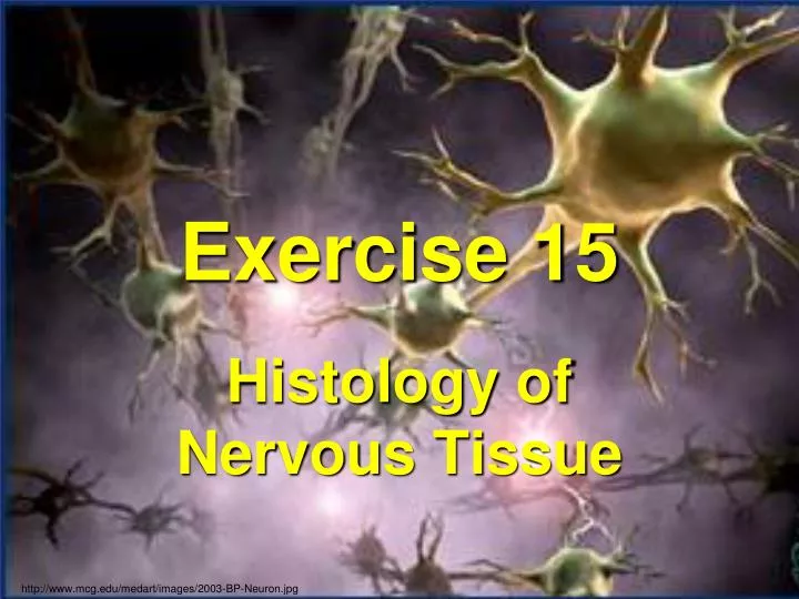
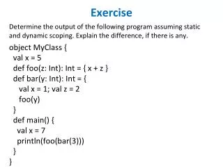
Exercise 15. Histology of Nervous Tissue. http://www.mcg.edu/medart/images/2003-BP-Neuron.jpg. (a) cell body (soma); (b) Nissl bodies; (c) dendrites; (d) axon; (e) axon hillock; (f) synaptic terminals (a.k.a., axon terminals).

E N D
Exercise 15 Histology of Nervous Tissue http://www.mcg.edu/medart/images/2003-BP-Neuron.jpg
(a) cell body (soma); (b) Nissl bodies; (c) dendrites; (d) axon; (e) axon hillock; (f) synaptic terminals (a.k.a., axon terminals). (a) Schwann cells; (b) neurilemma of a Schwann cell; (c) myelin sheath of a Schwann cell; (d) nucleus of a Schwann cell; (e) axon of a neuron; (f) node of Ranvier. (a) axon terminal; (b) synaptic cleft; (c) postsynaptic membrane; (d) synaptic vesicles (if shown)
Two Cell Types in Neural Tissue • Neuron • Neuroglia (glial) cell Fig. 12-1
Neuroglia (glial) cell • Several types Old Edition: 12-6
Capillary Neuron Astrocyte Figure 15.1 Neuroglia. Astrocytes are the most abundant CNS neuroglia Myelin sheath Process of oligodendrocyte Neuron Microglial cell Nerve fibers Oligodendrocytes have processes that form myelin sheaths around CNS nerve fibers. Microglial cells are defensive cells in the CNS. Fluid-filled cavity Cell body of neuron Cilia Satellite cells Schwann cells (forming myelin sheath) Ependymal cells Brain or spinal cord tissue Nerve fiber Ependymal cells line cerebrospinal fluid-filled cavities. Satellite cells and Schwann cells (which form myelin) surround neurons in the PNS.
CNS vs. PNS • Central Nervous System • Brain & spinal cord • Peripheral Nervous System • Cranial nerves, spinal nerves, ganglia, and nerve plexuses
Neurons classified by Function • Sensory (afferent) • Motor (efferent)
Figure 15.7 Classification of neurons on the basis of function. Ganglion Peripheral process (axon) Cell body Sensory neuron Central process (axon) Spinal nerve Afferent transmission White matter Interneuron Receptive endings Gray matter Efferent transmission Motor neuron Spinal cord (central nervous system) To effectors (muscles)
Neurons classified by Impulse Direction • Mixed • Carries sensory & motor • All spinal nerves, most cranial nerves • Pure • Carry only sensory or only motor Impulse toward CNS only (some cranial nerves) Impulse to an organ, muscle, etc. (some parts of spinal cord…)
Neuron Terminology • Ganglion • Cluster of cell bodies in the PNS • “gray matter” • Nuclei • Clusters of cell bodies in the CNS • Tract • Neuron processes in the CNS • “white matter” • Nerve • Neuron processes in the PNS
Figure 15.2a Structure of a typical motor neuron. Dendrites Cell body Nucleus Impulse direction Axon Myelin sheath gap (node of Ranvier) rough endoplasmic reticulum Axon terminals Axon hillock Schwann cell Terminal branches
Figure 15.2b Structure of a typical motor neuron. Nucleus of neuroglial cell Neurofibril Nucleus Nucleolus Dendrites Chromatophilic substance
Myelinated Nerve Fibers of PNS • Schwann Cells: • Neurilemma: peripheral part of cell • Nucleus: within the neurilemma (superficial) Fig. 12-5
Figure 15.3a Myelination of a nerve fiber (axon) by Schwann cells. (1 of 4) Schwann cell plasma membrane 1 A Schwann cell envelops an axon. Schwann cell cytoplasm Axon Schwann cell nucleus 2 The Schwann cell then rotates around the axon, wrapping its plasma membrane loosely around it in successive layers. 3 The Schwann cell cytoplasm is forced from between the membranes. The tight membrane wrappings surrounding the axon form the myelin sheath. Myelin sheath Schwann cell cytoplasm
Myelinated Nerve Fibers of PNS • Myelin sheath • Axon • Node of Ranvier Fig. 12-5
Synapse • Connection between the axonterminal & the next cell (presynaptic neuron) (postsynaptic neuron or other cell) Fig. 12-5
Figure 15.2c Structure of a typical motor neuron. Presynaptic neuron Direction of action potential Mitochondrion Synaptic cleft Axon terminal Synaptic vesicles Postsynaptic neuron
Synapse 12-2 • Synaptic vesicles • In axon terminal • Contain neurotransmitters • Acetylcholine most common Fig. 12-5
12-2 Nerve impulse travels Fig. 12-5
Structure of a Nerve 13-6
Figure 15.8a Structure of a nerve showing connective tissue wrappings. Axon Myelin sheath Endoneurium Perineurium Epineurium Fascicle Blood vessels
Figure 15.6 Photomicrographs of neurons. Dendrites Dendrites Cell body Cell body Nerve fibers Satellite cells Cell bodies

![[Exercise Name] Functional Exercise](https://cdn0.slideserve.com/621913/exercise-name-functional-exercise-dt.jpg)


![[Exercise Name] Functional Exercise](https://cdn1.slideserve.com/1717560/exercise-name-functional-exercise-dt.jpg)


![[Exercise Name] Functional Exercise](https://cdn3.slideserve.com/6680259/exercise-name-functional-exercise-dt.jpg)


![[Exercise Name] Tabletop Exercise](https://cdn4.slideserve.com/9191716/exercise-name-tabletop-exercise-dt.jpg)